From tissue uniformity to volume imaging
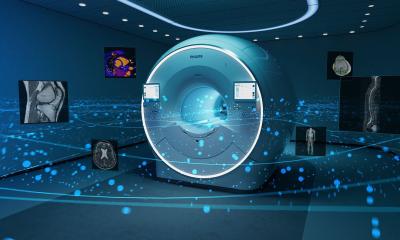
The latest breakthrough in premium ultrasound from Philips
Explaining the value of the iU22 xMATRIX ultrasound system, Philips describes its new array transducer within the system – the X6-1 PureWave xMATRIX – as revolutionary.‘It harnesses the power of over 9000 active elements, more than 35 times greater than conventional transducers, to capture crisp, high-resolution images of even technically challenging patients,’ the company points out, adding: ‘The X6-1’s next-generation beamformer technology features ultra-thin slice imaging that extends across the entire range to reduce artifacts and deliver exceptional quality 2D images. It offers extraordinary tissue uniformity for improved texture pattern resolution, as well as clear discrimination among near, mid, and far microstructures – to reveal anatomical detail never before seen.’

The X6-1 PureWave xMATRIX transducer features xPlane, which allows imaging in two planes without moving the transducer. The clinician therefore no longer has to rotate the transducer to see the second plane, or risk losing a tiny object during manual rotation. He gets twice as much clinical data in the same amount of time, allowing him to make the diagnosis faster, with increased confidence.
Last, new X6-1 PureWave xMATRIX transducer provides high resolution for both 2D and 3D imaging. As a result, there is no need to change transducers to acquire volume images, so there is no disruption to the examination. In fact, workflow is improved. And now, for first time ever, ultrasound volume data from any volume transducer is available on any PACS (DICOM multiframe object standard presently required for all cineloop information). Once the volume data is acquired, the iU22 will capture the X, Y, and Z MPR cineloops at the push of a button, and send them to the PACS. Simon Elliott, Consultant Radiologist and Clinical lead in ultrasound at Freeman Hospital, Newcastle upon Tyne, England, whose work is in abdominal, Doppler and small parts ultrasound, has worked with the xMATRIX ultrasound transducer for a couple of years. ‘I’m particularly interested in evaluating how this new technology could change our approach to diagnostic ultrasound,’ he explained, adding that radiologists are keen on CT and MR, but many patients are not so keen. A combination of the safety, speed, low cost and real-time imaging of ultrasound, and the benefits of CT/MR, that situation could be eased. ‘The xMATRIX transducer allows me to do this, by imaging conventionally in high resolution, or by acquiring 3D volumes at the touch of a button, plus other unique capabilities,’ Dr Elliott says.
‘The technology behind this development is astonishing. Instead of a single row of around 128 elements in a conventional transducer, the X6-1 xMATRIX transducer has over 9,000 elements arranged in a true matrix. Uniquely,’ Dr Elliott explains, ‘this allows for dynamic focusing in the elevation plane, as well as the standard azimuth plane, giving a very fine beam profile. Being electronic, the operator can select single or multiple planes or volumes while scanning in real-time. With so much processing involved, most of the beam-forming takes place in the transducer itself in a complex collection of integrated circuits containing more than eight million transistors.’ xPlane is particularly useful to obtain rapid coverage of the target organ or pathology with minimal physical effort, such as the gallbladder, kidney or urinary bladder, Drvolume estimations using this technique. But by utilising the full volume capability of the xMATRIX, we can begin to see how we approach the CT/MR model of working – and much faster.’
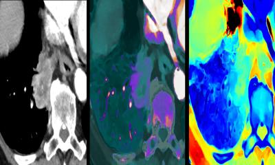
The X6-1 xMATRIX transducer can acquire the 3D shape of a full 90 by 90 degree volume, typically in under a second. ‘This means that you often don’t even have to ask the patient to stop breathing, which is particularly useful in the seriously ill patient, the elderly or in paediatrics. And, of course, this can all be acquired at the bedside.’ Among the examples Dr Elliott can present for the volume acquisition ultrasound Elliott adds: ‘We can perform rapid threediameter use, which include a simple renal cyst as well as a complex group of pancreatic pseudocysts, he describes an image of a portal vein in a liver transplant patient. Although it looks like a conventional 2D image, this is a selected plane from a half-second volume scan through the porta hepatis. The radiologist explains that he can go into that volume, locate the vessel and rotate the plane, capture the image and measurements wanted – all after the scan, just like CT/MR.’
For Dr Elliott, the xMATRIX X6-1 transducer has enabled him to perform many of the tasks normally associated with CT and MR imaging, ‘but much more conveniently for both me and the patient. It offers me speed, accuracy and flexibility in the way I use ultrasound in daily practice.’
“Advances in Ultrasound Using iU22 xMatrix Technology”, Philips Lunch Symposium,
Sunday, August 28, 12:15 – 13:15, Hall G
26.08.2011