Listen to your heart 24 hours a day
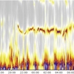
The ‘Fire of Life’ developed by Schiller is an intuitive visual presentation of frequency-domain heart rate variability (HRV) that makes the assessment of 24-hour results fast and simple.


The ‘Fire of Life’ developed by Schiller is an intuitive visual presentation of frequency-domain heart rate variability (HRV) that makes the assessment of 24-hour results fast and simple.

Carestream is receiving orders from healthcare providers around the world for its new Carestream Managed Print Solutions (MPS), a comprehensive, web-based, pay per print programme that tracks laser imaging film usage, and remotely monitors and delivers film inventory according to each facility’s needs. This comprehensive programme saves both time and money for healthcare providers.
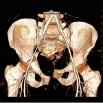
At this year’s meeting of the German Radiological Society (DRK), Dr Mathias Langer, Head of the Radiology Clinic at Freiburg University Hospital and the society’s 2013 President, assured EH that CT is still the be all and end all in trauma surgery.

Efforts to unify training and certification, a regulatory environment conducive to innovation and a growing bank of clinical evidence for key procedures, is helping interventional radiology (IR) to move to a new level.
Beckman Coulter, an indirect wholly-owned subsidiary of Danaher Corporation (NYSE:DHR), announced today that it has entered into a definitive agreement to purchase the clinical microbiology business of Siemens Healthcare Diagnostics.
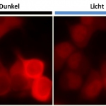
Researchers from the University of Heidelberg and the German Cancer Research Center (DKFZ) have developed a new method that uses light to control processes in living cells.

Agfa HealthCare announces today that it has signed an agreement with Real Time Medical (RTM) to support the needs of collaborating or consolidated hospitals and imaging networks.
COCIR, that represents the European Medical Imaging, Electromedical and Healthcare ICT Industry, announces the preliminary results of its Medical Imaging Equipment Age Profile and Density report.
Medicine as a profession has held a superior aloofness for many centuries, wary of losing its unique distinctiveness and esteem if ‘tainted’ with other professions.
About four years ago, Samsung Electronics Co. – specialist in electronic components and mobile phone sets, was recognised by its revenues as the world’s largest IT company, displacing Apple Inc.

Royal Philips today announced that it has signed a partnership agreement with the Stockholm County Council (SCC) to jointly innovate in health care.

Bracco Imaging, a global leading company in diagnostic imaging announced the launch of MultiHance (active ingredient: adobenate dimeglumine) in Russia.
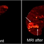
Over the next few years, the University Medical Center (UMC) Utrecht and Quirem Medical will be working closely together to maximize the benefits of using holmium microspheres to treat liver cancer patients worldwide.
MAQUET and Swisstom are pleased to announce their distribution partnership for the monitoring and diagnostic product Swisstom BB2.

The European Congress of Radiology (ECR), the biggest radiology meeting in Europe, was held March 6–10 in Vienna, Austria. Over 20,000 delegates from more than 110 countries attended the annual meeting of the European Society of Radiology (ESR), which took place for the 20th time at the Austria Center Vienna.

Claron Technology, the leader in universal medical imaging viewers, announces that it has been awarded the CE mark for its NilRead zero-footprint viewer for the diagnostic interpretation of medical images.
On March 6, COCIR* and ESR held a Joint Session at 2014 European Congress of Radiology (ECR) entitled “How does integrity affect our daily lives? A joint radiologist/industry initiative”.
Today, more than fifty percent of all ultrasound examinations are not performed by radiologists,” underlines Professor Dr Lorenzo Derchi, Head of Emergency Radiology at San Martino Hospital at University Hospital Genoa in Italy.
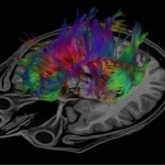
New insights into the ways the human brain functions – that is the promise of mapping the entire web of connections in the brain, the so-called connectome. New developments in connectome imaging are one of the major topics at this year’s European Congress of Radiology (ECR).
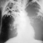
Approximately 1.7 billion people worldwide carry Mycobacterium tuberculosis (MTB), i.e. about one-third of the population. The rate of new infections is highest in Africa, followed by certain Asian regions, including areas in Russia.
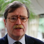
Two things that radiologists resist – structured reporting and (computer-assisted) quantification – are the very things that Gabriel Krestin believes are essential to advance diagnosis in the brave new world of omic-medicine that is emerging.

Nothing is more personal than a scan of the insides of your body. Yet radiology images today look a little old-fashioned and out-of-step in the fast-emerging movement to personalised medicine.
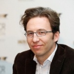
‘Elastography is in a position much like Doppler 20 years ago,’ according to David Cosgrove, BMBCh, MA, FRCR, FRCP, Professor of Clinical Ultrasound at Imperial College School of Medicine in London.
Workstations and desktops may still be around in future hospitals – will be of clear benefit to medics in a large variety of medical applications.
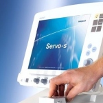
Toshiba’s new 1.5-T MRI Vantage ELAN system is not only cost-effective, the firm reports, but truly compact; it needs only 23 square metres of space. Yet, the system uses the same type of magnet as other Toshiba products to achieve excellent image quality.