Image source: Radboudumc
News • Position and completeness assessment
3D CT analysis benefits liver tumor ablation
Important step towards broader use of ablation treatment
By analyzing CT images with 3D software, small liver tumors can be successfully treated using ablation. This has been demonstrated by research conducted at Radboud university medical center (Radboudumc). These findings, published in CardioVascular and Interventional Radiology, enable more confident use of ablation in treating patients.
Liver tumors up to three centimeters can be treated in various ways. In consultation with the patient, the treating physician chooses between surgery or ablation, a method in which the tumor is punctured and heated with a thin needle until it is destroyed. The latter method is less invasive; it is quicker, carries a lower risk of complications, and allows for faster recovery. Interventional radiologist Sjoerd Jenniskens and technical physician Kristian Overduin jointly perform this procedure at Radboudumc. Overduin explains: ‘Ablation offers many benefits for the patient. Until now, however, it has been difficult to assess whether all tumor cells have been treated.’
This is an important step toward broader application of ablation for small liver tumors. For patients, it means lower risk of complications and faster recovery
Kristian Overduin
Oncological surgeon Martijn Stommel shares this view. ‘We’ve been working closely with our radiologists for years to make optimal use of imaging in the treatment of liver tumors. The risk of cancer recurrence was relatively high with ablation compared to surgery. By jointly reviewing our methods and the available technical possibilities, we developed a new process,’ says Stommel.
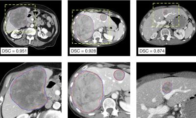
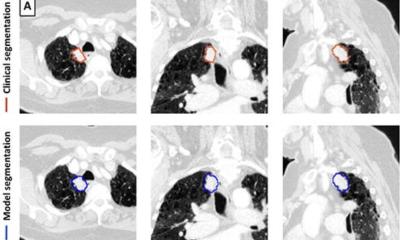
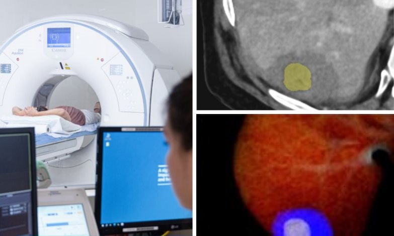
A key element of this new process is 3D analysis. The ablation does not take place in an operating room, but in a room equipped with a CT scanner. Immediately after the procedure, while the patient is still under anesthesia, a CT scan is taken. “Previously, this scan was assessed visually. Now we use 3D software for the analysis, providing a much clearer image of the treatment result. We overlay pre- and post-treatment scans and can instantly see whether the entire tumor has been treated. If not, we can continue the procedure immediately,” Overduin explains. He led a study evaluating the outcomes of this new process.
The results are promising. Thanks to the 3D software analysis, the recurrence rate of liver tumors within two years after ablation has dropped significantly—from 33% to less than 10%. Moreover, patients required fewer repeat treatments. “These numbers are very impressive,” says Overduin. “This is an important step toward broader application of ablation for small liver tumors. For patients, it means lower risk of complications and faster recovery.”
“The 3D analysis is a great example of smart use of technology that directly benefits the patient,” says Overduin, who is also part of Radboudumc’s Talent Track (intranet), the talent program for exceptionally talented young scientists. “With this innovation, we contribute to affordable healthcare. Less invasive procedures benefit patients but also reduce costs and ease the burden on healthcare systems. All of this is the result of close collaboration between interventional radiologists, oncological surgeons and hepatologists. Together with other Dutch liver expert centers. It’s gratifying to make a meaningful contribution.”
Source: Radboud university medical center
29.07.2025