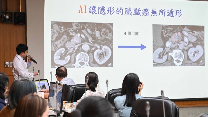
Image source: NTUH
News • Diagnostic assistance
Pancreatic cancer: clinical deployment of AI system for early detection
Pancreatic cancer is one of the deadliest and most challenging malignancies worldwide due to its asymptomatic onset, diagnostic difficulty, and rapid progression.
It is projected to become the second leading cause of cancer-related death in the United States by 2030, and has already risen to seventh place among cancer-related deaths in Taiwan. The overall five-year survival rate remains around 10%, but this could increase to 80% if tumors smaller than 2 centimeters are detected and treated early. However, such early-stage pancreatic tumors are notoriously difficult to spot on CT images, with approximately 40% of lesions missed. Diagnosis remains heavily reliant on the experience and capacity of radiologists.
In response to this challenge, National Taiwan University Hospital (NTUH), in collaboration with the Institute of Applied Mathematical Sciences at National Taiwan University, has developed the world’s first AI-powered pancreatic cancer diagnostic system: “PANCREASaver”. Officially launched in late 2024 at NTUH’s Department of Medical Imaging, this tool is now available as a self-pay AI image analysis service, marking a major milestone in the early detection and precision treatment of pancreatic cancer.
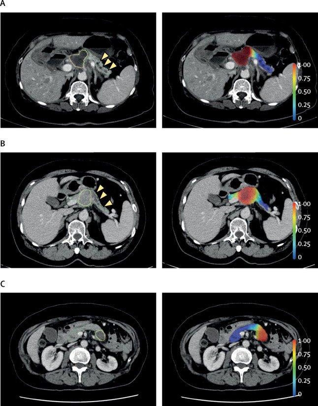
Image source: Liu KL, Wu T, Chen PT et al., Lancet Digital Health 2020 (CC BY 4.0)
The pancreas lies deep within the abdominal cavity, making its tumors difficult to identify via conventional CT imaging—especially those under 2 cm, which are often imperceptible to the human eye. PANCREASaver addresses this diagnostic gap by leveraging deep learning algorithms and multi-image training data to automatically analyze CT scans, delineate the pancreas, and flag suspicious lesions directly within the hospital’s PACS system.
National clinical validation trials have demonstrated that PANCREASaver achieves an 80% sensitivity rate for early-stage pancreatic tumors (<2 cm) and an overall diagnostic accuracy exceeding 90%, significantly enhancing radiologists’ confidence and efficiency in making timely diagnoses.
PANCREASaver is now fully deployed at NTUH, allowing radiologists to instantly access AI-generated results to support more efficient decision-making. The system is also integrated into multidisciplinary care, including gastroenterology, surgery, and oncology, enabling personalized and timely treatment strategies. In one clinical case, a 67-year-old female patient presented with jaundice and itching. Her initial CT scan revealed dilation of the common bile duct and intrahepatic bile ducts, but no visible pancreatic mass. With the help of PANCREASaver and an endoscopic ultrasound (EUS), a hidden lesion was identified in the pancreatic head. The diagnosis of early-stage pancreatic cancer was confirmed, allowing prompt surgical treatment.
PANCREASaver has been awarded four invention patents in Taiwan and four in the United States, and has received regulatory approval from the Taiwan Food and Drug Administration (TFDA) as well as the U.S. FDA Breakthrough Device Designation. Its research outcomes have been published in journals such as The Lancet Digital Health and Radiology. The system has also received multiple honors, including the RSNA Margulis Award, National Innovation Award, Taipei Biotech Award, and the SNQ National Quality Certification.
The PANCREASaver development team aims to expand its applications to other pancreatic-related conditions, such as pancreatitis and pancreatic cystic lesions, while incorporating multi-modal imaging and clinical data to support broader risk assessment and precision treatment strategies. This new AI model not only solidifies NTU Hospital’s leadership in AI-integrated medical imaging but also establishes a new global benchmark for early pancreatic cancer detection, offering new hope and life-saving opportunities for patients worldwide.
Source: National Taiwan University Hospital
23.05.2025