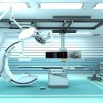
Photo

News • Gathering sonography experience

Partnership to provide ultrasound online interactive training

Royal Philips and LeQuest, a provider of online interactive simulation-based training for the use of medical devices, announced a partnership to provide online interactive training for the Philips Ultrasound Affiniti system. As a result, medical staff can train individually at their own convenience without the need for equipment, avoiding the resulting loss of operational time on the system, as…

Photo

News • Technologies combined

New fluoroscopy system combines fluoroscopy and radiography

Siemens Healthineers is debuting its new fluoroscopy system, the Luminos Lotus Max, at the annual French JFR 2020 (Journées Francophones de Radiologie Diagnostique & Interventionnelle). The system offers industry-leading technology in radiography and fluoroscopy. “Combining these state-of-the-art technologies poses one major challenge: ensuring efficient workflows and high system…

Photo

News • Cardiovascular Magnetic Resonance

Insight on the use of CMR in COVID-19 patients

The Society for Cardiovascular Magnetic Resonance (SCMR), a Society dedicated to improving cardiovascular health by advancing the field of cardiovascular magnetic resonance (CMR), has released a formal statement supporting and explaining their position on the use of CMR in COVID-19 patients. The topic has led to debate among medical professionals, especially those in the cardiology specialty.

Photo

Article • Modality comparison

COVID-19 imaging: lung ultrasound vs chest CT

A recent preprint study in France underpins the debate on whether lung ultrasound (LUS) should be used to triage COVID-19 patients better at the hospital as well as in primary care. The eChoVid study, published as a preliminary report of work on medTrix, shows that LUS enables identification of lung lesions as well as chest CT in COVID-19 patients. A team of French researchers compared routinely…

Photo

Article • POCT as initial coronavirus screening tool

Ultrasound confirms frontline value in COVID-19 setting

Ultrasound could become the prime modality in emergency settings for tracking disease progression in COVID-19 patients. While chest CT has held a key diagnostic role thus far, many experts now advocate the benefits of ultrasound within the context of the coronavirus epidemic. Dr Rachel Liu, who recently led a high-profile panel discussion with experts from the USA and areas of Europe with high…

Photo

News • Tiny chip, huge benefits

Researchers develop the world’s smallest ultrasound detector

Researchers at Helmholtz Zentrum München and the Technical University of Munich (TUM) have developed the world’s smallest ultrasound detector. It is based on miniaturized photonic circuits on top of a silicon chip. With a size 100 times smaller than an average human hair, the new detector can visualize features that are much smaller than previously possible, leading to what is known as…

Photo

Article • Non-pulmonary aspects

The cardiovascular impact of Covid-19

The increased Covid-19 risk to cardiac patients was discussed during an online presentation at ECR 2020. Focusing on the prevalence of pulmonary embolism in Covid-19 patients, Dr Karl-Friedrich Kreitner, Professor of Radiology at the Department of Diagnostic and Interventional Radiology at the Johannes-Gutenberg University in Mainz, Germany, discussed hypotheses which can explain cardiac…

Photo

News • head and neck radiology

ESHNR 2020 Online Teaching Weeks

ESHNR is proud to bring you 20 hours of online education in the form of on-demand webinars. For a period of 6 weeks (October 24 – December 7, 2020), registered attendees can log in and watch at their own discretion a careful selection of webinars on the most common head and neck topics.

Photo

News • Outstanding radiologist

International recognition for Dr Jamie MacKay

Radiologist Dr Jamie MacKay has achieved international recognition for his research using advanced imaging to help patients with osteoarthritis and other musculoskeletal conditions. MacKay, who is a radiologist at the Norfolk and Norwich University Hospital (NNUH) and lecturer at University of East Anglia (UEA), has been elected as a Junior Fellow of the International Society for Magnetic…

Photo

Video • Multiple sensors to capture high-res images

A new detector for long-length digital radiography

Carestream Health has introduced a new digital radiography detector in the United States and Canada, enabling hospitals and imaging centers that specialize in long-length imaging to increase productivity while decreasing both patient discomfort and dose. With a single exposure, the Carestream DRX-L Detector captures long-length images by providing a large field of view and high resolution for…

Photo

Article • Neuroradiology

Alzheimer’s research: A lost century

Lack of understanding around Alzheimer’s disease (AD) has significantly slowed advances in the treatment of this incurable condition. Imaging has proved to be reliable in differentiating between AD and other forms of dementia, and its contribution will continue to help develop profiling, an increasingly interesting approach for the development of new and more efficient drugs, according to Sven…

Photo

Article • Neuro-oncology

Challenges in brain tumour segmentation

Neuroradiologist Dr Sofie Van Cauter described the challenges to brain tumour image segmentation during the European Society of Medical Imaging Informatics (EuSoMII) annual meeting in Valencia. She also outlined how, when clinically validated, AI could help tackle such problems. The WHO classification of brain tumours has come a long way since first introduced in 1979. The 2016 classification was…

Photo

News • Showcased at virtual ISMRM meeting

MRI image analysis and workflow software platform launched

Random Walk Imaging AB (RWI), a company developing novel software solutions for diffusion magnetic resonance imaging (MRI), announced the launch of its first commercial software product for clinical researchers and radiologists. The dViewr Powered by Mice Toolkit is the result of a collaboration with Nonpi Medical AB, with whom RWI has entered into an exclusive license and development agreement…

Photo

News • Improved accuracy and efficiency

AI could improve CT screening for COVID-19

Researchers at the University of Notre Dame are developing a new technique using artificial intelligence (AI) that would improve CT screening to more quickly identify patients with the coronavirus. The new technique will reduce the burden on the radiologists tasked with screening each image. Testing challenges have led to an influx of patients hospitalized with COVID-19 requiring CT scans which…

Photo

News • POCUS equipment

New point-of-care ultrasound system for better workflow

Fujifilm Sonosite, Inc. has announced the launch of the new Sonosite PX ultrasound system. It is the next generation in the manufacturer's point-of-care ultrasound (POCUS) devices, with the most advanced image quality ever seen in a Sonosite system, a suite of workflow efficiency features, and an adaptable form factor.

Photo

Video • Improving first aid

New 3D CT scanning method shows what happens during CPR

Rapid first aid during cardiac arrest makes the difference between life and death. But what happens to the heart and the internal organs when people come running and begin to give well-meaning but heavy-handed heart massage as they attempt to keep the person who has suffered a cardiac arrest alive? A research collaboration between the Department of Forensic Medicine at Aarhus University and the…

Photo

News • Breast cancer imaging

Should Gadolinium-based contrast agents be used in breast DTI?

The accuracy of breast cancer diagnosis via diffusion-tensor imaging (DTI) was equivalent both before and after the administration of a gadolinium-based contrast agent (GBCA), despite a value change in DTI parameters, finds a new research article. “However,” wrote first author Anabel M. Scaranelo of Princess Margaret Cancer Centre’s breast imaging division in Toronto, “the limitations in…

Photo

Article • Future of contrast agents

Gadolinium in MRI is here to stay (at least for a while)

Manganese and iron oxide contrast agents can replace gadolinium-based contrast agents (GBCA) in a number of MRI examinations, but gadolinium remains a strong candidate when properly indicated, especially with AI-driven dose reduction and advances to increase relaxivity, a French expert explained at ECR 2020. GBCA have been MRI companions for many years. In France, 30% of all MR examinations are…

Photo

News • Corona in radiology

Revealing COVID-19-related brain injury in MRI and CT imaging

Injuries in the nervous system of patients with severe COVID-19 are revealed by magnetic resonance imaging (MRI) and computed tomography (CT). In a study of 185 patients, researchers at Karolinska Institutet (KI) and Karolinska University Hospital show an affection of microscopic blood vessels and inflammation in the brain, meninges and nerves. The results are published in Radiology.

Photo

Sponsored • After the earthquake

Ultrasound provides much-needed answers for rebuilding lives in Nepal

Dr. Jesus Casado Cerrada, Internist at the Hospital Universitario de Getafe and Professor at the Universidad Europea, Madrid, Spain, has travelled to the Rasuwa district of Nepal to help a local NGO rebuild the region’s infrastructure following a severe earthquake in 2015. Dr. Casado explains: “University colleagues from the architectural and engineering departments had already established…

Photo

News • Deep learning in imaging

1.5T MR system receives FDA clearance for AI-based image reconstruction technology

Canon Medical Systems USA, Inc. has received 510(k) clearance on its Advanced intelligent Clear-IQ Engine (AiCE) for the Vantage Orian 1.5T MR system, continuing to expand access to its new Deep Learning Reconstruction (DLR) technology. This technology, which is also available on the Vantage Galan 3T MR system and across a majority of Canon Medical’s CT product portfolio, uses a deep learning…

Photo

News • Nanoparticles

Luminescent substance allows accurate viewing of body parts

Researchers have designed a fluid that works like a luminous ink to obtain very sharp images of damaged tissues, organs and cartilages in diagnostic tests. This new compound, still in the laboratory phase, reduces adverse effects on the human body because it allows lower amounts to be injected and the dose to be targeted only at the affected area.

Photo

News • 3D microprinted scope

World’s smallest imaging device focuses on heart disease

A team of researchers led by the University of Adelaide and University of Stuttgart has used 3D micro-printing to develop the world’s smallest, flexible scope for looking inside blood vessels. The camera-like imaging device can be inserted into blood vessels to provide high quality 3D images to help scientists better understand the causes of heart attack and heart disease progression, and could…

2392 show more articles
Subscribe to Newsletter