Sponsored • Patient positioning
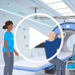
Relief for radiographers: The Get Up holding system for back-friendly repositioning
Discover in only one minute, how the Get Up holding system sustainably assists both patients and medical staff.


Discover in only one minute, how the Get Up holding system sustainably assists both patients and medical staff.
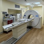
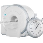
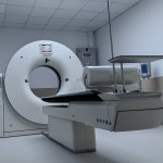
Siemens Healthineers expands its 1.5 Tesla Magnetom Flow. Platform to include a model with a 70-centimeter bore with the introduction of a new magnetic resonance imaging (MRI) scanner at RSNA 2024.
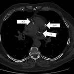
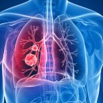
As new research shows, low-dose chest computed tomography (CT) can identify coronary artery calcium, a strong risk factor for coronary artery disease (CAD), in patients without cardiac symptoms.
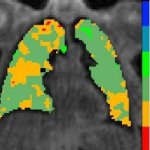
After a Covid-19 infection, patients may suffer from a variety of symptoms, including difficulty concentrating (“brain fog”). New research now linked the condition to impaired lung function.
United Imaging are excited to share that, in partnership with Fora S.p.A., an industry leader with over 50 years of expertise in delivering advanced diagnostic technologies to the Italian market, Policlinico Casilino Hospital will join the company's global network. Demonstrating its commitment to innovation and growth, the hospital has selected the industry-leading 640-slice CT scanner to…
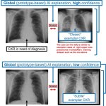
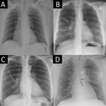
When an AI advisor points out an area of concern in a chest X-ray, radiologists are sometimes all too eager to follow their lead, a new study finds. This may lead to incorrect diagnostic decisions.
Royal Philips announced a major advance in radiation oncology with 510(k) clearance from the US Food and Drug Administration (FDA) for its new detector-based spectral CT radiotherapy solution.

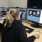
Radiology AI products are a whole new world. So is running them safely and efficiently in production. At the 2024 Society for Imaging Informatics in Medicine (SIIM) Annual Meeting, an expert outlined how new skills and resources are needed to use these AI tools – and how PACS administrators could evolve their position to fit this important role.
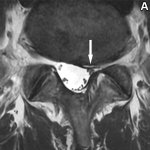
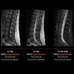
Not every spine abnormality shown in an MRI scan causes pain for a patient. To diagnose correctly, researchers advocate the use of questionnaires to match the images with reported symptoms.
Because radiology exams are an integral part of the treatment process for many hospital inpatients, any improvements in efficiency can have a positive ripple effect on routine hospital operations and functionality. Two quality control presentations at the 2023 RSNA Annual Meeting shared the theme of creating representational staff teams to identify bottlenecks involving radiology report…
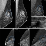
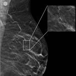
Evidence of the superiority of tomosynthesis for breast cancer detection is stacking up, with new results from a 10-year study further demonstrating the 3D imaging technique's benefits.
Interdisciplinary collaboration between gynaecologists, radiologists, pathologists and breast care nurses following a palpation finding makes a decisive contribution to the success of further breast cancer treatment. This was the consensus among the speakers at the Annual Congress of the German Society of Senology in Dresden. The experts provided clear explanations of which imaging is best for…
Radiology practices have a high volume of chest X-rays without clinically significant finding, which take up a lot of time. A new AI tool could improve workflows by providing an automatic report.
Artificial Intelligence will be a critical component in ensuring a Europe-wide lung cancer screening programme can achieve its potential, according to speakers at a special ECR 2024 session. Delegates heard that the SOLACE project (Strengthening the screening of Lung Cancer in Europe) will be supported by AI in terms of workflow, diagnostics, and image and data analysis.
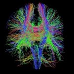
Just a concussion? Head injury can lead to persistent symptoms, yet CT scans often fail to identify these cases. Researchers explore the potential of diffusion tensor imaging for better diagnostics.

As the adoption of AI into healthcare continues, concerns about the environmental impact of increasingly complex AI models grow. Therefore, researchers are looking for more sustainable AI solutions.
As opportunities for teleoperations rapidly expand within radiology, the concept is being deployed across an array of modalities to deliver more efficient healthcare. A range of speakers covered the topic of ‘Teleoperations in radiology’ at ECR2024, discussing its benefits in applications in MRI, ultrasound, during the social restrictions of the Covid-19 pandemic and military use. However,…

Using an advanced scanner, researchers have developed a technology that can detect the earliest changes in the kidney when scar tissue begins to form.
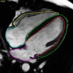
From 45 minutes to just a few seconds: A new computer model utilises AI to examine cardiac MRI scans in the four-chamber plane, potentially offering speedy and dependable heart health evaluation.
Using specific radiomics features from 70 characteristics in MRI images, researchers develop an objective method to predict the hearing status of patients with vestibular schwannoma.
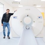
United Imaging announces that it has provided the Unicamed Hospital in Gjilan, Kosovo, with two state-of-the-art systems – the uCT 528 80-slice CT scanner and the uDR 266i digital radiography system.

Digital twin technology can transform clinical practice by aiding patient-specific prediction and supporting personalized treatment models. Expert speakers at an ECR 2024 session in Vienna focussed on how radiology will play a leading role in the advance through data acquisition via a range of imaging modalities.
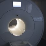
The IRCCS in Bologna has inaugurated a state-of-the-art integrated PET/CT system. This cutting-edge technology allows for the entire human body to be studied in a single scan, even detecting the smallest tumour cells.
Through the joint efforts of United Imaging and NeuralMed, Prizren Regional Hospital has become the first facility in Kosovo to benefit from United Imaging's cutting-edge diagnostic technology.
Current AI systems for detecting breast cancer from mammography exams are more likely to produce false-positive results in black women and older patients, a new study finds.