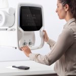
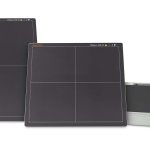
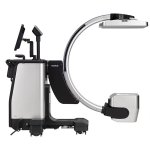
News • NewTom 7G
The next level of 3D X-ray imaging
Precision, speed and simplicity of use. Safety for patient and X-ray technician. Reduced total cost of ownership. These are the benefits of the first Multi-Scan Body CBCT developed by NewTom.
In radiology, it is important to place patients safely and precisely. This works with the "GetUp®" holding system from Febromed.
A new study has found that AI can help physicians in interpreting x-rays after an injury and suspected fracture. This has the potential to improve diagnostic performance and efficiency.
Scientists have used tin mono-sulfide (SnS) nanosheets to create the thinnest X-ray detector ever made, potentially enabling real-time imaging of cellular biology.
The damage caused by Covid-19 to the lungs’ smallest blood vessels has been intricately captured using high-energy X-rays emitted by a special type of particle accelerator. Scientists used a new imaging technology called Hierarchical Phase-Contrast Tomography (HiP-CT), to scan donated human organs, including lungs from a Covid-19 donor.
A team of scientists at Argonne National Laboratory has leveraged artificial intelligence to train computers to keep up with the massive amounts of X-ray data taken at the Advanced Photon Source.
At a virtual European event, Fujifilm Healthcare Europe presented a complete and integrated portfolio of diagnostic products and services, including CT, MRI, X-ray, AI, PACS, endoscopy and ultrasound systems. This launch follows the completion of Fujifilm's acquisition and takeover of Hitachi's Diagnostic Imaging-related business on 31 March 2021 for 179 billion yen (€1.3 billion).
University of Washington researchers have discovered that AI models—like humans—have a tendency to look for shortcuts. In the case of AI-assisted disease detection, these shortcuts could lead to diagnostic errors if deployed in clinical settings.
Before the huge potential of tiny nanocarriers for highly targeted drug delivery and environmental clean-up can be realized, scientists first need to be able to see them. Currently researchers have to rely on attaching fluorescent dyes or heavy metals to label parts of organic nanocarrier structures for investigation, often changing them in the process. A new technique using chemically-sensitive…
Carestream Health has released Smart Noise Cancellation (SNC), a groundbreaking artificial intelligence (AI)-based technology that greatly improves image quality — producing images that are significantly clearer than with standard processing.
Kobe University Hospital’s Dr. Makoto Nishimori and Project Assistant Professor Kunihiko Kiuchi et al. (of the Division of Cardiovascular Medicine, Department of Internal Medicine) have developed an AI that uses multiple kinds of test data to predict the location of surplus pathways in the heart called ‘accessory pathways’, which cause the heart to beat irregularly. In this study, the…
At this year's virtual ECR congress, Ziehm Imaging showcases its portfolio of mobile C-arms and advanced imaging solutions. Highlights on display include two clinical packages for cardio and vascular interventions based on the fully motorized Ziehm Vision RFD Hybrid Edition, that provide clinical users with state-of-the-art imaging support and further workflow integration. The virtual booth,…
Clinicians will be able to take a low radiation, digital X-ray image - without the need for an anti-scatter grid - thanks to new innovative ‘scattering suppression software’. Developed by photonics scientists at the Warsaw University of Technology (WUT), working in collaboration with innovation incubator ACTPHAST 4.0 and medical imaging company Italray SRL, the new algorithm ‘auto…
Displaying medical images from different modalities such as CT, CR/DR, MR, ultrasound or mammography and pathology on one monitor - that is what most radiologists want. Many of them still use several medical displays next to each other. JVCKenwood's new CL-S1200 30.9-inch colour monitor makes this a thing of the past. The 12 megapixel device (4,200 horizontal and 2,800 vertical) can display…
Hologic, Inc. has announced it has CE Mark for the Faxitron Path+ Specimen Radiography System, the latest addition to the company’s growing portfolio of digital specimen radiography solutions. The Faxitron Path+ system features a superior, high-resolution imaging detector which enables pathologists to image a wide range of specimens of varying sizes, including bone, foetal remains and breast…
In clinical practice, planar X-ray images are usually the first imaging technique used. In musculoskeletal imaging, this can be a shoulder in AP and lateral projection. In many of these cases, the 2D technique is not sufficiently informative and further clarification is required, which means additional waiting times or completely new appointments. The DTS allows the diagnostic cycle to be…
Researchers have designed a fluid that works like a luminous ink to obtain very sharp images of damaged tissues, organs and cartilages in diagnostic tests. This new compound, still in the laboratory phase, reduces adverse effects on the human body because it allows lower amounts to be injected and the dose to be targeted only at the affected area.
Computed tomography (CT) is one of the most important imaging options for the diagnostics of patients. For a long time, in clinical routine CT technology mostly showed the Hounsfield Units (HU) that are indicated in the grayscale. However, the Dual Energy CT (DECT) enables colored X-ray images, therefore significantly improving examination methods. “We can also do material separation and get a…
Welcome to the world premiere of YSIO_x.pree.
Researchers at the University of Zurich have developed a new X-ray contrast agent which is easier to use and distributes into all blood vessels more reliably, increasing the precision of vascular imaging. This reduces the number of animals required in research experiments. Various diseases in humans and animals – such as tumors, strokes or chronic kidney disease – damage the blood vessels.…
The organ of balance in the inner ear is surrounded by the hardest bone in the body. Using synchrotron X-rays, researchers at Uppsala University have discovered a drainage system that may be assumed to play a major role in the onset of Ménière's disease, a common and troublesome disorder. These results are published in the journal Scientific Reports. Ménière's disease is manifested in sudden…
Although the clinical symptoms of new pediatric lung disorders such as severe acute respiratory syndrome (SARS), swine-origin influenza A (H1N1), Middle East respiratory syndrome (MERS), e-cigarette or vaping product use-associated lung injury (EVALI), and coronavirus disease (COVID-19) pneumonia may be nonspecific, some characteristic imaging findings "have emerged or are currently…
Hsinhan Tsai and a research team in materials, nanotechnology, nuclear engineering and X-ray science at the Los Alamos National Laboratory and the Argonne National Laboratory in the U.S. demonstrated a new thin film X-ray detector prototype.
A new X-ray detector prototype is on the brink of revolutionizing medical imaging, with dramatic reduction in radiation exposure and the associated health risks, while also boosting resolution in security scanners and research applications.
Like most technologies, X-ray imaging software is evolving, delivering more automation and improved image quality. However, the fundamental imaging basics for radiologic technologists remain unchanged. In this article, manufacturer Carestream provides a helpful review of the imaging basics for rad techs. In today’s digital world, basic functions once performed by the operator are now…