Image source: Pyrros et al., Nature Communications 2023 (CC BY 4.0)
News • Deep learning in imaging
Earlier detection of diabetes through chest x-rays and AI
A new artificial intelligence (AI) model finds that x-ray images collected during routine medical care can provide warning signs for diabetes, even in patients who don’t meet the guidelines for elevated risk.
The model could help physicians detect the disease earlier and prevent complications, says a multi-institutional team led by Emory University which published the findings in Nature Communications.
Applying the computational method known as deep learning to images and electronic health record data, the researchers developed a model that successfully flagged elevated diabetes risk in a retrospective analysis, often years before patients were diagnosed with the disease. That’s significant, the researchers say, given the prevalence of diabetes in the U.S. has more than doubled over the past 35 years.
Recommended article

News • From 529 million to 1.3 billion cases by 2050
Diabetes: Experts predict drastic increase in the next 30 years
More than half a billion people are living with diabetes worldwide, and that number is projected to more than double to 1.3 billion people in the next 30 years.
Current guidelines suggest screening patients for type 2 diabetes if they are between 35 and 70 years old and have a body mass index (BMI) in the overweight to obese range. Many studies, however, have found this strategy misses a significant number of cases, particularly in racial/ethnic minorities for whom BMI is a less effective predictor of diabetes risk. Patients with undiagnosed diabetes are at much higher risk for complications from the disease including irreversible organ damage and even death.
Chest x-rays provide an ‘opportunistic’ alternative to universal diabetes testing. This is an exciting potential application of AI to pull out data from tests used for other reasons and positively impact patient care
Judy Wawira Gichoya
Each year, millions of Americans receive chest x-rays for chest pain, difficulty breathing, injury or before surgeries. Emory alone completes on average about 200,000 radiographs annually. While radiologists are not looking for diabetes when they assess these x-rays, the images become part of a patient’s medical record and could be analyzed later for diabetes or other conditions. “Chest x-rays provide an ‘opportunistic’ alternative to universal diabetes testing,” says Judy Wawira Gichoya, MD, assistant professor of radiology and imaging sciences, and the lead researcher from Emory. “This is an exciting potential application of AI to pull out data from tests used for other reasons and positively impact patient care.”
The AI model was trained on more than 270,000 x-ray images from 160,000 patients, with deep learning determining the image features that best predicted a later diagnosis of diabetes. Because chest x-rays are not a common way to detect diabetes, the researchers also used explainable AI techniques to determine how and why the model made its determinations. The methods pointed to the location of fatty tissue as important for determining risk, a logic that aligns with recent medical findings that visceral fat in the upper body and abdomen is associated with type 2 diabetes, insulin resistance, hypertension and other conditions.
Given the novelty of the approach and the startling results, the researchers who first developed the model turned to the Emory team to perform external validation of the model. When the Emory team applied the model to a separate group of nearly 10,000 patients, they found the model predicted risk better than a simple model based on non-image clinical data alone.
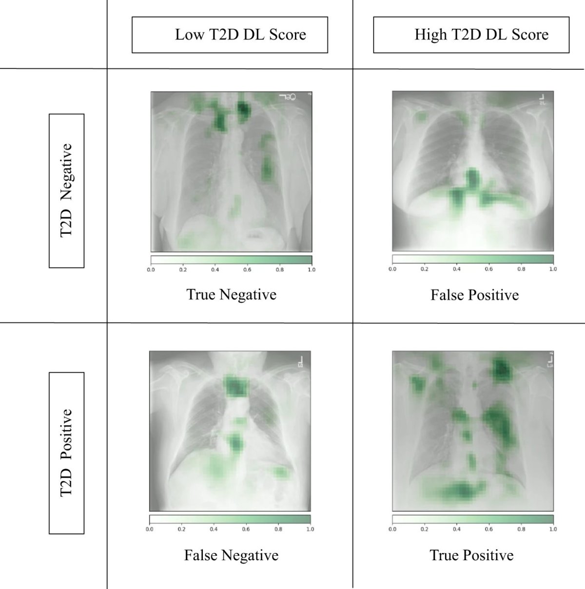
Image source: Pyrros et al., Nature Communications 2023 (CC BY 4.0)
In some cases, the chest x-ray warned of high diabetes risk as early as three years before the patient eventually received a diagnosis. The model’s output also provides a numerical risk score that could potentially help clinicians customize the treatment approach for patients.
“Diabetes is a chronic disease where body fat distribution matters. The longer the duration of the disease and the worse the glycemic control, the higher the risk of complications,” says Francisco Pasquel, MD, associate professor in Emory’s Division of Endocrinology, Metabolism and Lipids. “The opportunistic approach of using chest x-rays to identify those at the highest risk of diabetes, even before a spike or drop in blood sugar levels occurs, is a promising method that may help improve outcomes through early preventive measures or treatment.”
The research team will now explore how to further validate the model and incorporate it into electronic health record systems so it can provide an alert to physicians to pursue traditional diabetes screening of patients flagged as high risk based on x-ray results. They’ll then turn to investigating how well chest x-rays can help diagnose other conditions, such as vascular disease, congestive heart failure, and chronic obstructive pulmonary disease (COPD).
The research was primarily funded through a grant from the NIH Medical Imaging and Data Resource Center.
Source: Emory University
04.08.2023