Credit: Newcastle University, UK
News • Bacterial protection strategy
Protective 'chain mail' of C.difficile revealed
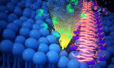
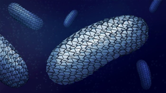
The spectacular structure of the protective armour of superbug C.difficile has been revealed for the first time showing the close-knit yet flexible outer layer – like chain mail. This assembly prevents molecules getting in and provides a new target for future treatments.
The scientists from Newcastle, Sheffield and Glasgow Universities together with colleagues from Imperial College and Diamond Light Source, outline the structure of the main protein, SlpA, that forms the links of the chain mail and how they are arranged to form a pattern and create this flexible armour. This opens the possibility of designing C. diff specific drugs to break the protective layer and create holes to allow molecules to enter and kill the cell.
One of the many ways that diarrhoea-causing superbug Clostridioides difficile has to protect itself from antibiotics is a special layer that covers the cell of the whole bacteria - the surface layer or S-layer. This flexible armour protects against the entry of drugs or molecules released by our immune system to fight bacteria.
The team determined the structure of the proteins and how they arranged using a combination of X-ray and electron crystallography.
“I started working on this structure more than 10 years ago, it’s been a long, hard journey but we got some really exciting results! Surprisingly, we found that the protein forming the outer layer, SlpA, packs very tightly, with very narrow openings that allow very few molecules to enter the cells. S-layer from other bacteria studied so far tend to have wider gaps, allowing bigger molecules to penetrate. This may explain the success of C.diff at defending itself against the antibiotics and immune system molecules sent to attack it," said corresponding author Dr Paula Salgado, Senior Lecturer in Macromolecular Crystallography who led the research at Newcastle University.
“Excitingly, it also opens the possibility of developing drugs that target the interactions that make up the chain mail. If we break these, we can create holes that allow drugs and immune system molecules to enter the cell and kill it,” she added.
One of the current challenges in our fight against infections is the growing ability bacteria have to resist the antibiotics that we use to try to kill them. Antibiotic or more generally, antimicrobial resistance (AMR), was declared by WHO as one of the top 10 global public health threats facing humanity.
Different bacteria have different mechanisms to resist antibiotics and some have multiple ways to avoid their action – the so-called superbugs. Included in these superbugs is C. diff, a bacteria that infects the human gut and is resistant to all but three current drugs. Not only that, it actually becomes a problem when we take antibiotics, as the good bacteria in the gut are killed alongside those causing an infection and, as C. diff is resistant, it can grow and cause diseases ranging from diarrhoea to death due to massive lesions in the gut. Another problem is the fact that the only way to treat C.diff is to take antibiotics, so we restart the cycle and many people get recurrent infections.
Determining the structure allows the possibility of designing C. diff-specific drugs to break the S-layer, the chainmail, and create holes to allow molecules to enter and kill the cell.
Colleagues, Dr Rob Fagan and Professor Per Bullough at the University of Sheffield carried out the electron crystallography work: “We’re now looking at how our findings could be used to find new ways to treat C. diff infections such as using bacteriophages to attach to and kill C. diff cells - a promising potential alternative to traditional antibiotic drugs,” he said.
From Dr Salgado’s team at Newcastle University, PhD student Paola Lanzoni-Mangutchi and Dr Anna Barwinska-Sendra unravelled the structural and functional details of the building blocks and determined the overall X-ray crystal structure of SlpA. Paola said: “This has been a challenging project and we spent many hours together, culturing the difficult bug and collecting X-ray data at the Diamond Light Source synchrotron.”
Dr Barwinska-Sendra added: “Working together was key to our success, it is very exciting to be part of this team and to be able to finally share our work.”
The paper was published in Nature Communications.
Source: Newcastle University
27.02.2022