News • MedTech
Concerning fall in UK radiology equipment spend
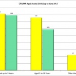
The overall UK radiology equipment market spend for the six months to the end of March 2018 is down by around 30% compared to the same period in the previous year. This is according to latest figures from AXREM (the Association of Healthcare Technology Providers for Imaging, Radiotherapy and Care), which represents all the major medical imaging manufacturers active in the UK. Commenting on the…