News • Brain imaging
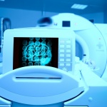
FDA approves first neonatal MRI
The U.S. Food and Drug Administration cleared the first MRI device specifically for neonatal brain and head imaging in neonatal intensive care units.


The U.S. Food and Drug Administration cleared the first MRI device specifically for neonatal brain and head imaging in neonatal intensive care units.

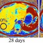
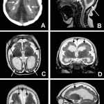
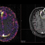
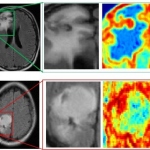
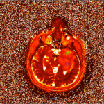
Scientists from the German Cancer Research Center (DKFZ), in collaboration with colleagues from Heidelberg University Hospital, have been able to visualize brain cancer using a novel MRI method. They use a simple sugar solution instead of conventional contrast agents, which can have side effects in the body.

Gadolinium-based contrast agents are an essential component of MRI exams, but are challenged by findings of residual depositions of gadolinium in the body, even though the clinical relevance remains unknown. Three clinicians described how changes to MRI protocols and dose levels for contrast media can optimise the balance between benefit and risk for patients and radiologists.
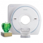
Toshiba Medical proudly announces its 2nd MRI User Meeting in collaboration with Clinica Creu Blanca in Barcelona on 22 & 23 September 2017 (at Camp Nou FC Barcelona, Spain). At this MRI User Meeting, international experts will share their experiences and clinical solutions for successful diagnostic imaging in Women's and Men's Healthcare.

MRI enhanced with gadolinium ethoxybenzyl diethylenetriamine pentaacetic acid -the scan referred to as “EOB MRI” - is significantly better than contrast-enhanced CT for assessing colorectal liver metastases that disappear after chemotherapy, according to a study published online March 22 in Radiology.
New MRI techniques are set to offer advances in the earlier detection of liver disease in patients. Radiologists are harnessing the potential of highly-targeted MRI, whilst exploring the imaging modality as a means of delivering non-invasive biomarkers, reducing the need for biopsy to measure treatment response.

The ProMRI Configurator made by Biotronik is an online tool that enables physicians to select from a series of MRI requirements for a patient and subsequently generates a recommendation of all suitable MR-conditional cardiac device and lead combinations available in a particular country, thus helping physicians to choose the most suitable MR-conditional cardiac systems for each patient.
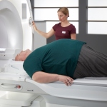
Just in time for the ECR, Siemens Healthineers is coming up with an innovation. Magnetom Vida, the new high-end 3 Tesla MRI scanner with BioMatrix technology from Siemens Healthineers, was launched to the public at University Hospital Tübingen yesterday, where the first system is installed. The new BioMatrix technology adapts automatically to individual anatomical and physiological…

What can we learn from population studies? According to Gabriel Krestin MD PhD there are things that we can un-learn, as well as learn, from population imaging studies.
ECR 2017 Guest Lecturer Maria de Fatima Vasco Aragao, a radiologist from Pernambuco state, Brazil, has been tracking the Zika virus ever since it broke out in her country in 2015. She will highlight how CT and MRI can help reach diagnosis, especially in the absence of microcephaly. In an exclusive interview with European Hospital correspondent Mélisande Rouger, the radiologist warned there might…
From the extremely new, but not very available, to the somewhat new, very available and highly useful, Walter Kucharczyk will cover the potentials and practicalities in advanced brain tumor imaging.

Have you seen the videos? Perhaps you have read the 20-page paper published in Radiology? At MR 2017 Garmisch you will have the chance to see and hear a live presentation by William Palmer, M.D., Director of Musculoskeletal Radiology & Intervention at Massachusetts General Hospital, a teaching hospital of the Harvard Medical School.
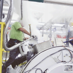
Helium, a critical component in MRI systems, has gone through two potential shortage crises, impacting hospitals and patients around the globe. But the helium supply is finite and demand has been rising over the past decades. At #RSNA16, GE Healthcare proudly unveils Freelium*, a magnet technology designed to use one percent of liquid helium compared to conventional MRI magnets. Instead of the…
‘Magnetic resonance imaging is a very dynamic field,’ declared Professor Siegfried Trattnig, head of the Centre of Excellence for High Field MRI in the Department of Biomedical Imaging and Image-guided Therapy, at Vienna Medical University. Indeed, this September, two mega trends emphasised by Trattnig – the shift from qualitative to quantitative imaging and Big Data – dominated the 33rd…
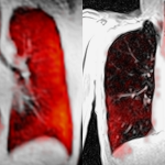
Since lung diseases tend to be complex, imaging is a crucial diagnostic tool. While computed tomography has become the standard modality, which is frequently used outside hospital settings, specialised MRI diagnostics remains the preserve of large university medical centres.
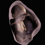
Parents may soon be able to watch their unborn babies grow in realistic 3D immersive visualizations, thanks to new technology that transforms MRI and ultrasound data into a 3D virtual reality model of a fetus, according to research being presented next week at the annual meeting of the RSNA.
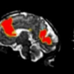
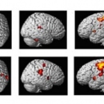
NIBIB-funded researchers at the University of Washington have pioneered an approach to image functional activity in the brains of individual fetuses, allowing a better look at how functional networks within the brain develop. The work addresses a common problem of functional MRI; if the subject moves during the scanning, the images get distorted.
Computer programs have defeated humans in Jeopardy!, chess and Go. Now a program developed at Case Western Reserve University has outperformed physicians on a more serious matter.
Bracco Imaging S.p.A., a global leading company in the diagnostic imaging business, today announced the approval of the use of MultiHance® (gadobenate dimeglumine) in Magnetic Resonance Imaging (MRI) of the whole body in adults and paediatric patients (> 2 years). The approval was obtained through a Mutual Recognition Procedure, with the Medicines and Healthcare products Regulatory Agency…
The 32nd Congress of the European Committee for Treatment and Research in Multiple Sclerosis opened in London (September 14-17) and key presentations reveal the latest evidence-based thinking on how to use MRI scan results to diagnose MS with increasing accuracy. Earlier this year, MAGNIMS, the European Collaborative Research Network that studies the use of MRI when diagnosing MS, published…
From the beginning of MRI scientists dreamed of not only translating data sets into images, but also classifying tissue directly. Five years ago this would still have been inconceivable.
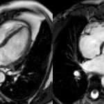
Scientists have shown that people who exercise for even a few hours each week can enlarge their hearts. This is a normal and beneficial response to exercise, but until now has only been recognised in athletes. The researchers say that doctors should now consider an individual’s activity level before diagnosing common heart conditions.
UK researchers are working on a more precise imaging technique for dilated cardiomyopathy that may lead to more effective treatments. A study from the University of Oxford Centre for Clinical Magnetic Resonance Research (OCMR), part of the Division of Cardiovascular Medicine at the university, has demonstrated how the next generation of MRI scanners can work to measure heart conditions in dilated…
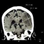
Stroke patients will first undergo a CT scan as they enter the hospital. Before any further imaging scan is carried out, the medical team must decide whether they need to intervene intra or extra cranially. ‘Imaging enables you to see which pathology you are dealing with and helps you select patients for either recanalisation or revascularisation or, in some cases, occlusion by embolisation,’…
MRI increasingly helps to diagnose cardiac disease, yet its role in clinical decision-making of acutely hospitalised patients has hardly been explored.