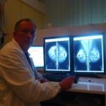
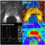
Radiology & nuclear medicine - Sharing an awkward waltz in Vienna
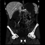
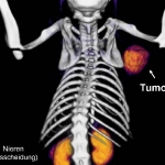
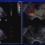
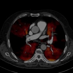
CT-PET is the child of a forced marriage between nuclear medicine and radiology. A shared session at ECR 2011 in Vienna did little to assure there is a growing consensus between the two partners.