Does a foetus think - if so, how?
MRI in foetal screening
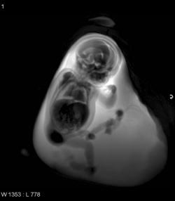
Ultrasound is the undisputed choice for foetal imaging. However, the lack of amniotic fluid, an unfavourable foetal position or maternal obesity could daunt even the best radiographer. In such circumstances prenatal magnetic resonance imaging (MRI) – a non-invasive and non-irradiating procedure – is a helpful diagnostic tool.
It is also considered if the ultrasound image does not offer conclusive findings, e.g. an unclear malformation. ‘Or,’ as Dr Daniela Prayer, paediatric radiologist at the University Clinic for Radiological Diagnostics, Vienna University Hospital, put it, ‘if there is uncertainty regarding normal organ development. Or if a malformation is detected that is often associated with a genetic syndrome and it needs to be confirmed or excluded. A further indication for prenatal MRI is the presence of familial genetic defects, which means the imaging procedure aims to check whether the foetus shows morphology that indicates such a defect. Unclear miscarriages in the mother’s family may also indicate prenatal MRI.’
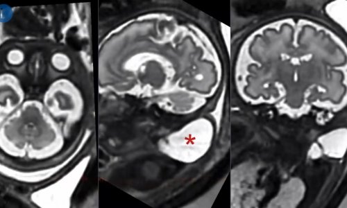
Almost all foetal body parts (e.g. face, neck, thorax, abdominal organs, surplus renal tissue) as well as maternal tissue can be reliably and precisely visualised in prenatal MRI. Brain development can be assessed with almost histological precision. Detection of the intrinsic movements of organs in real time is also possible. ‘If a foetus with a stenosis in its oesophagus has problems swallowing amniotic fluid,’ Dr Prayer explained, ‘this can be detected with the help of prenatal MRI, which enables obstetricians to plan for possible post-partum surgical procedures.’
The good news for clinical practice is that high quality prenatal MRI does not necessarily require state-of-the-art technology, e.g. a 3-Tesla or even 7-T system. 1.5-T can yield very good results, Dr Prayer believes. Whilst she uses 3-T for research she said, ‘If and when the 3-T will dominate foetal imaging is entirely unclear. But we expect to be able to answer this open question in about half a year.’
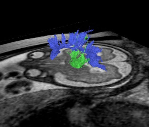
MRI applications continue to expand, she said. ‘We are looking at morphology, but recently functional MRI has been allowing us to visualise connectivities in the brain, to observe non-brain organ development and to assess metabolic processes.’
As early as the 17th or 18th week of pregnancy, Dr Prayer performs resting state examinations to assess brain activity. This procedure will in the medium term allow categorisation of normal and deviant brain activities.
Initial results should be ready for publication in early 2011.
28.10.2010