Article • MRI
9-, 10- or even 11-Tesla – is more always better?
Advancing magnetic resonance imaging (MRI) up the Tesla scale may sound good, but will it produce the results and patient safety radiologists actually desire?
Faced with the question: ‘How many Tesla should it be?’, Professor Siegfried Trattnig MD, head of the Centre of Excellence in high-field MRI at the University Clinic for Radiodiagnostics, Medical University of Vienna, and Austria’s only professor of high-field MRI research, provided his answers at the Austrian-Bavarian Radiology Congress held in Linz this October.
The 1.5 Tesla MRI scanner, according to Professor Siegfried Trattnig, is still the ‘workhorse’ of radiology, but this, he foresees, may change in just a few years. ‘The trend with new installations is definitely heading towards 3-T scanners.’
Today, some 15 years after the first generation of high field strength MRI was introduced, the advantages are quite clear: better image quality and quicker examination times, which means higher patient capacity. Economic aspects also make 3-T MRI more interesting for private practices. 3-T scanners have also proved themselves in research around functional MRI (fMRI). ‘For example, the equipment is used in our brain activity studies and for metabolic examinations,’ Prof Trattnig pointed out. ‘Whereas we only capture about 2-3% of the so-called image interferences that occur due to paramagnetic haemoglobin with our 1.5-T scanner, 3-T scanners capture 4-6% of these interferences. And now we even have 7-T scanners that actually allow us to capture up to 10% of interferences.’
High-field MR imaging

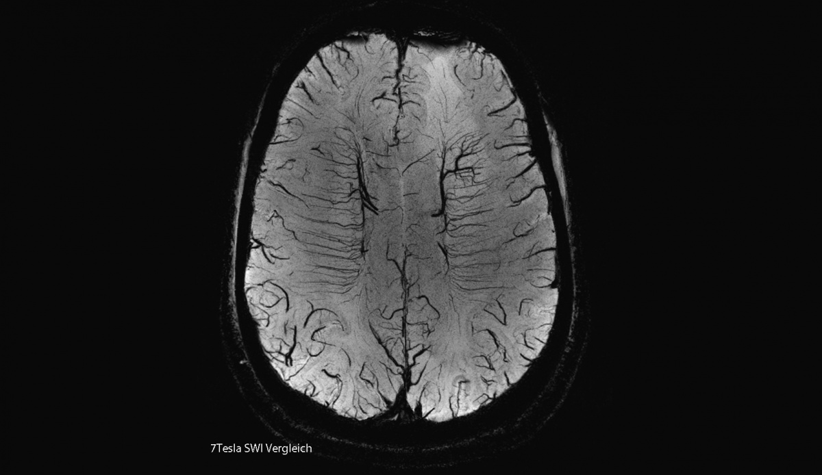
Worldwide, thirty-five 7-Teslas have been installed, and purely for research – none have entered clinical routine. With good reason, he believes. ‘With 7-T scanners we must work very methodically to compensate for the disadvantages of stronger artefacts. With this high field strength we also encounter inhomogeneities of the magnetic field, which we must first get to grips with so that we can then actually utilise the advantages of high field strength at all. Additionally, although a 3-T scanner is delivered complete with all protocols, when a 7-T scanner acquisition the coils must be specifically developed and manufactured separately. So these scanners are currently only used in combination with hardware development, fundamental research, method development and clinical studies. Basically, there’s a development process between science and industry before the equipment will become deployable for future, practical use.’
That future may not be so distant. Medical University of Vienna studies have shown that functional MRI (fMRI) with 7-T is more precise in pre-operative planning for brain tumour treatment than scanners with lower field strengths. This is also something appreciated by neurosurgeons at the AKH Vienna, who want this technology established in clinical practice. ‘In some sub-specialties, such as neurology, the advantages of 7-T are already so significant that, in certain cases, we should not stop the technology being offered to patients,’ Prof. Trattnig believes, although exclusively for use in large clinical centres; he sees no need for the use of these strong magnets by private practice doctors.
We can only guess the kind of physiological effects 11.4 Tesla scanners might have on people.
Siegfried Trattnig
With 7-T scanners heading for routine use, how far off might the routine use of the 11.4-T MRI scanner be? ‘A difficult subject,’ he acknowledged, because ‘the prestige project near Paris clearly shows the limitations of the technology. On the one hand there’s the structural implementation required to shield the magnetic field, which is very complex. It requires building elements from the field of nuclear fusion. On the other hand, occasionally 7-T scanners have already shown physiological effects on people – dizziness, a metallic taste or pulses of light before patients’ eyes. We can only guess the kind of physiological effects 11.4 Tesla scanners might have on people. Against this background, it’s probably right to say that the technology will take us toward the limitations of its possible, practical use in human medicine. Considering the technical complexity and costs, as well as additional gain in information, in my opinion only a field strength up to a maximum of 9- or 10-T will make sense for radiological diagnosis in human medicine.’
Profile:
Siegfried Trattnig MD became Medical Head of Austria’s first high-field (3 Tesla) MRI research scanner ten years ago and, in 2003, founded the Centre of Excellence for High-Field Magnetic Resonance Imaging at the Medical University of Vienna. Today, as Professor for Radiology and specialist in high-field MRI, he heads the 7-T and 3-T Project at the university. The professor has pioneered parametric or biochemical imaging of articular cartilage with MRI sequence and procedure development, up to its clinical use after cartilage replacement.
28.10.2010