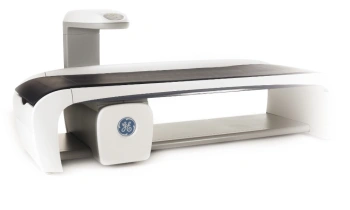
An improved view with 64 slices
Meike Lerner reports
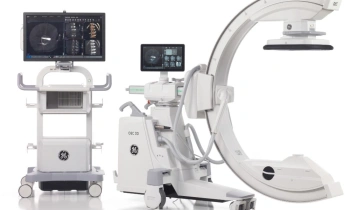
Late in 2009, GE Healthcare’s 64 slice PET/CT Discovery 690 began its use in the University Clinic for Nuclear Medicine in Innsbruck, Austria. Apart from routine clinical examinations for treatment control via FDG (fluorodeoxyglucose), the scanner is also employed in clinical research - particularly for Gallium 68DOTA PET/CT examination.
‘We are a worldwide leader in this field,’ Professor Irene Virgolini, Head of the University Clinic, pointed out. ‘We’ve already supported many institutions in establishing Gallium 68 examinations.’
The procedure is particularly used in cases of neuro-endocrine tumours and radioiodine-negative thyroid carcinoma, where it is mainly used for process monitoring of high-dose treatment. ‘We were significantly involved in the development of the generator that generates the Gallium 68, and we produce various compounds, so-called analogues, which are then used for PET/CT diagnostics,’ said Prof. Virgolini. ‘Currently, for example, we are in the process of developing a specific liver tracer with a Gallium 68 marker, which can then show liver reserve capacity. At the moment this is a market niche, which we hope to fill. We also have a tracer for angiogenesis imaging, which is already undergoing clinical studies. It is aimed at patients who take an angiogenesis inhibitor. We have high hopes that this tracer could have significant savings potential, if it enables us to determine the therapy response.’
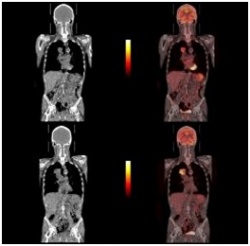
A further important field is viability determination in cardiac examinations with unclear results. According to Professor Virgolini, the strength of the PET/CT here lies in the ability to display a clearer image of perfused areas with non-perfused areas due to its better resolution, analogue to the accumulation of FDG. Even though nuclear medicine has always delivered very good results in this field, the combination with a 64-slice CT is advantageous due to the image overlay.
In general, the Innsbruck Clinic has been very pleased with this new technology. ‘One of the advantages is that the scanner is a lot faster, and the exposure to radiation is also lower,’ Prof. Virgolini observed. ‘Our objective for the current year is to carry out 3,500 examinations - clinical examinations as well as research-related ones.’
However, there is one downer, which also affects Austria: Medical insurers continue to refuse reimbursement of the FDG examinations. Prof. Virgolini: ‘In the German speaking countries we are significantly behind in this field, even though the latest developments point towards a need for change in reimbursement policies.’
08.09.2010
- CT (617)
- display (438)
- imaging (1676)
- medical technology (1580)
- neurology (639)
- nuclear medicine (132)
- PET/CT (192)