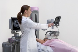
The automated volume breast scanner
Ultrasound catches up with slice imaging
Innovative technologies such as elastography, shearwave and 3-D have been cause for euphoria in today’s ultrasound world. The most recent development in breast imaging also has the potential to revolutionise this field, because the Automated Volume Breast Scanner (ABVS) is allowing user-independent and reproducible image acquisition for the first time.
Thus, ultrasound has finally caught up with other diagnostic imaging modalities and can be integrated with modern multi-modality concepts used in breast diagnostics. Dr Frank Stöblen, diagnostic radiology specialist and co-owner of Diavero, a diagnostic imaging centre in Essen, Germany, has worked with the Siemens Automated Breast Volume Scanner Acuson S2000 ABVS for over a year. As the first European user to have performed more than 2,000 examinations with this new technology, he described his experiences and observations in an interview with Daniela Zimmermann of European Hospital.
ABVS solves many problems of ultrasound technology in general, said Dr Stöblen. ‘First, the technology is automated, which means the transducer, user-independently, always generates images of the same quality. While ultrasound is one of the most frequently used imaging technologies, it is often done incorrectly and the technology itself, as well as its application, is subject to very few quality assurance checks. Thus, hand held ultrasound is an extremely subjective procedure with results entirely dependent on the knowledge and experience of the physician.
‘ABVS, however, scans the entire breast automatically without user intervention. Moreover, integrated 3-D technology provides coronal views that were impossible to generate with conventional ultrasound equipment. The physician no longer has to be present during ABVS procedures. The examination can be performed by assistants. Ultrasound has always been a very haptic technology and therefore I’m sure it will take some time before radiologists get used to the new concept. Working with ABVS is a bit like driving a remote-controlled car.
‘The second advantage is the reproducibility of full-field volume images. ABVS generates a complete data set that can be called up on a workstation – wherever and whenever needed. Sure, in the past ultrasound images were integrated into the PACS, but these were always a few isolated images. Now we can store up to 1,800 images per examination and scroll through them like we scroll through a video – just the way it’s done in multislice CT. This
has diagnostic potential, because we can now detect architectural deformations that we couldn’t see before. Furthermore, a second opinion can now be based on the exact same images as the primary opinion. ‘And, last but not least, research and training will benefit from the volume data sets as they allow a complete retrospective analysis and can be used as teaching files.’
When do you use ABVS?
‘In early detection ultrasound improves tumor detection above all in women with dense breast tissue. We were able to show in some smaller studies that ABVS can find all solid lesions. In a cancer prevention study with 300 previously examined women, with an average age of 30 years, ABVS detected two carcinomas.
‘As far as benign lesions are concerned, 3-D ultrasound is even slightly better than mammography. It can help to clarify ambiguous findings in dense tissue, such as cysts. However, ultrasound is not suited to detect microcalcifications.
Will there be additional applications beyond breast imaging?
‘Definitely. I am sure there will be applications for other organs, suchas liver, vessels and the thyroid gland, very soon. Mamma diagnostics was just the beginning. It allowed us to gather know-how since the breast is a surface organ and easily accessible. Moreover, mamma diagnostics is moving towards multi-modality. You do need all three modalities – mammography, ultrasound and MRI – in order to perform a differential diagnosis. Consequently, there are more and more multi-modality workstations in which the ultrasound images can now be integrated – thanks to the volume scanner.
What technological enhancements do you expect for ABVS?
‘The next step will be the integration of elastography into the 3-D system. Most likely shearwave will be the best technology, but at this point it cannot be realized since during tissue measurement with elastography the region of interest must not move -- a tricky technological problem but not impossible to solve.’
07.09.2010