Real-time tissue elastography as a complementary procedure
Tissue hardness provides radiologists and gynaecologists with significant information to help distinguish between benign and malignant tumours. Tumour tissue is harder and less malleable than normal glandular and fatty tissues. Therefore, the classification of tissue hardness determines whether a biopsy is necessary. For breast diagnoses, real-time tissue elastography, along with conventional B-image ultrasound, helps to improve diagnostic specificity through tissue hardness analysis.

In Germany, a multicentre study has released important findings on the suitability of sonoelastography in clinical practice. Involved in the study, Dr Thomas Fischer, at the Institute for Radiology, Charité Berlin and Professor Friedrich Degenhardt, Senior Consultant at the Gynaecology and Obstetrics Clinic in Franziskus Hospital Bielefeld, discussed the study findings with European Hospital.
The value of previous sonoelastography studies has involved only small cohorts of patients. At the Charité Berlin, in Bielefeld-Herford and in Bad Homburg, data from 779 patients was evaluated. Following the discovery of a lesion, each received an ultrasound-guided punch biopsy and analysis if their tissue samples then determined whether elastography delivers better results than B-image ultrasound. The data evaluation showed that, for certain subgroups, ‘it is imperative that sonoelastography is carried out,’ concluded Prof. Degenhardt. ‘This includes patients with breast cancer in BI-RADS categories three and four. In these cases, sonoelastography allows for a much more explicit differentiation between benign and malignant lesions.’
Sonoelastography has a specificity of 89.5% compared to 76.1% for conventional B-image; the positive predictive value can also be significantly increased from 77.2% to 86.6%. For focal findings in the BI-RADS categories III and IV diagnostic accuracy improves significantly by using elastography and the data also shows improved specificity of 92.8% in the case of dense glandular parenchyma.
The data have resulted in a current study group recommendation to the American College of Radiology to include sonoelastography in the BI-RADS atlas, so that it can be used in a similar way to the power Doppler or the colour Doppler as a complementary procedure. ‘Our objective is to see this procedure being used purposefully and comprehensively to help either to prevent a biopsy in the case of a clearly benign diagnosis or, in the case of an ambiguous diagnosis, to ensure that biopsies are definitely carried out if a lesion has been assessed as malignant based on the elastography,’ Dr Fischer explained, and, Prof. Degenhardt added: ‘We obtain the morphological information through the B-mode of the ultrasound and can gather additional, important information about the hardness of these lesions with elastography.’
Many medical technology manufacturers have now integrated elastography software into their ultrasound scanners as an additional option. The differences in the quality of the technology depend less on the elastography tool than on the B-image quality, Dr Fischer emphasised: ‘If I don’t find a lesion during the first step then I have nothing to carry the elastography out on. Therefore elastography will only ever be as good as B-image ultrasound implemented in the equipment, which is what points towards a suspicious area in the first place.’
Dr Fischer recommends that new, interesting developments, such as sheer wave elastography, should always be assessed as part of the whole picture: How good is the B-image? How good is the quality of elastography? How does the evaluation tool work? The improvement of primary B-mode screening is also close to his heart because it can spare patients an MRI scan, which some find claustrophobic, and a biopsy is also quite complex in the narrow tube. ‘Although MRI is supersensitive, unfortunately it is also unspecific. However, despite this problem, we depend on MRI in certain cases to find certain types of lesions. Preliminary stages of breast cancer and invasive lobular carcinoma can be captured well by MRI, in their entirety. However, once they have been detected, we can then often also see them on the ultrasound and can then determine whether the tumours are benign or malignant via punch biopsy,’ he pointed out. Elastography can also be used in a helpful way for this ‘second look’.
* Multicentre Study of Ultrasound Real-Time Tissue Elastography in 779 Cases for the Assessment of Breast Lesions: Improved Diagnostic Performance by Combining the BI-RADS(R)-US Classification System with Sonoelastography. Wojcinski S, Farrokh A, Weber S, Thomas A, Fischer T, Slowinski T, Schmidt W, Degenhardt F.
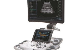
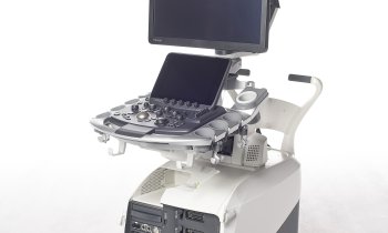
The HI VISION Preirus by Hitachi Medical Systems provides:
● Second generation graphical user interface with Smart Touch Technology
● Patient Scanning Selector (PSS)
● New image formats
● Patient Dependent Correction (PDC)
● Real-time Tissue Elastography (HI-RTE)
●Dynamic Contrast Harmonic Imaging (dCHI)
●Real-time Virtual Sonography (RVS)
●Biopsy Guidance
●Single Crystal Transducer Technology
●Full DICOM connectivity
03.11.2010


