Video • New treatment options
Unlocking the secrets of cell communication
Portland State University (PSU) researchers have made a significant breakthrough by developing the 3-D structure of proteins from inside the eye lens that control how cells communicate with each other, which could open the door to treating diseases such as cataracts, stroke and cancer.
The PSU research team, led by chemistry professor Steve Reichow, used a multimillion-dollar microscope and a novel technique developed by three Nobel Prize-winning biophysicists called cryo-electron microscopy (Cryo-EM) to view membrane protein channels — or transportation tunnels in cell walls — at the atomic level.
This allowed Reichow’s team, whose research is supported by the National Institutes of Health, to create a 3-D image of the membrane channel to better understand the processes involved in cell-to-cell communication.
Our discovery may one day pave the way for development of pharmaceuticals that can control heart disease or other diseases that are associated with the malfunctions or mis-regulations of these protein channels
Steve Reichow
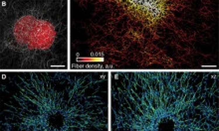
Portland State researchers used Cryo-EM — a microscope technique that freezes biomolecules in mid-movement and takes ultra-high-resolution images — and computer modeling to see the 3-D structure of gap junction proteins that had been isolated from eye lenses. Gap junctions are tiny channels that allow neighboring cells to communicate with one another and are found in many places throughout the body. Their findings — published Dec. 12 in the scientific journal Nature — showed for the first time how gap junctions selectively pass or block chemical information. Until now, it was not known how these channels would allow certain messages to pass between cells while specifically blocking others.
Reichow said the detailed images may open the door to understanding, and potentially treating, different types of diseases that are associated with the loss of function in cellular communication involving gap junctions such as cataracts, cardiac arrhythmia, stroke and certain cancers. "Currently there's no drug on the market today that can specifically block or activate gap junction proteins," Reichow said. “But our discovery may one day pave the way for development of pharmaceuticals that can control heart disease or other diseases that are associated with the malfunctions or mis-regulations of these protein channels."
Now that the Reichow Lab has established a method for characterizing the proteins, they are trying to understand the nuances of how the body uses gap junctions differently across tissues and organs.
Source: Portland State University
13.12.2018