Source: Philipp Brantner/3D-Printing Lab at the University Hospital Basel
Article • ECR 2019
The impact of 3D printing in radiology
With increased precision, speed of service and reduced cost, 3D printing presents an opportunity to transform traditional healthcare and its delivery, and radiology is at the center of this new technology. In the ECR 2019 Special Focus Session “The 3D printing lab from bench to bedside”, the speakers emphasized that 3D printing does not only enable a new and innovative way to display imaging, but it also contributes to patient care and allows radiologists to offer clinical value to their medical and surgical colleagues.
Report: Sascha Keutel
With 3D printing, the radiological workflow is changing dramatically, as it is moving from the traditional stepwise referral-report approach to a totally different device-oriented process, said Assoc. Univ.-Prof. Dr. Franz Kainberger, deputy chief of the Division of Neuroradiology and Musculoskeletal Radiology at the University Hospital Vienna. He therefore expressed the need for radiologists to define the role of 3D printing into their field. “This is a crucial topic of our work in the near future: if we want to include 3D printing into our daily work, we have to be aware that it is not the real image interpretation or diagnosis report in radiology. What we want to use is a printed diagnosis,” he said. “Especially in non-standardized procedure. That’s the real added value of some 3D models.” According to Kainberger, 3D printing should be regarded as a holistic approach that substantially influences the personalization of imaging diagnostics with a prognostic, a preventive and a participatory impact.
Source: Philipp Brantner/3D-Printing Lab at the University Hospital Basel
A 3D printing lab in radiology
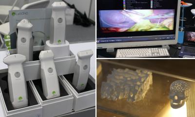
“3D printing is on the rise with increasing numbers of publications, interest from both industry and research in the fields of procedure planning, development of medical devices and implants, tissue engineering and education,” said Francesco Moscato, Associate Professor at the Center for Medical Physics and Biomedical Engineering, Medical University of Vienna.
But what is the magic of 3D printing, what makes it revolutionary? Moscato asked the audience. “It basically produces a physical model out of nowhere, out of a digital model. It is groundbreaking because it allows in a relatively simple process to produce geometrically complex and accurate models,” he said. “The key working principle is the automatically controlled layer-by-layer deposition of materials.”
Moscato went on to list the range of activities of a 3D printing lab in a clinical environment:
- The surgical and interventional procedure planning
- Creating models for training and simulation, but also functional models, e.g. for the cardiovascular field where you can get information about patient-specific fluid flow profiles
- Developing medical devices or improving existing ones
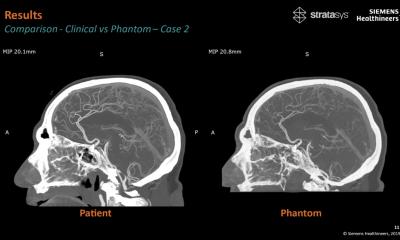
- Producing phantoms for clinical imaging optimization
Moscato explained that creating a 3D printing lab takes on the challenges of combining and coordinating knowledge from different technical and clinical disciplines such as referring physician, radiologists and engineers while at the same time guaranteeing strict adherence to medical regulations.
Moscato went on to explain the iron law of 3D printing: “your 3D model can’t be better than your medical image”. In general, the image quality should be as high as necessary, but according to him, there are conflicting views and tradeoffs:
Clinical:
- Reduction of patient burden: lower radiation dose and scanning time
- Lower image quality often sufficient for clinical diagnosis
- Human skills compensate for lower image quality
3D printing:
- Best possible image for intended use needs high radiation dose and scanning time
- Lower image quality often results in poor 3D models
- Rapid development of algorithmic compensation, but still far from human skills.
Source: Philipp Brantner/3D-Printing Lab at the University Hospital Basel
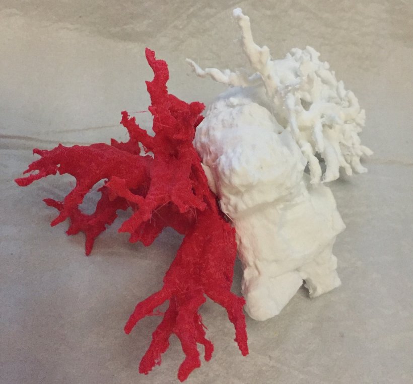
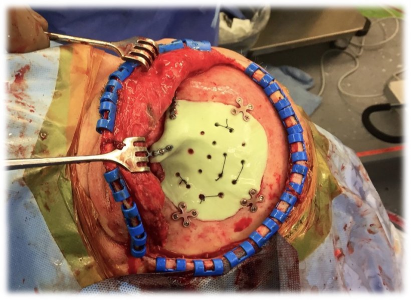
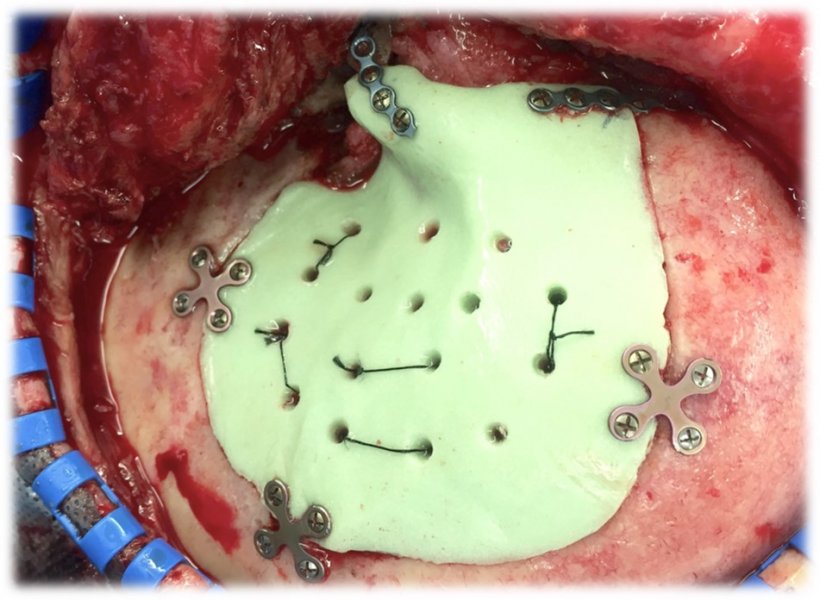
Supporting the surgeon with 3D printing
According to Dr. Philipp Brantner, Co-Director of the 3D-Printing Lab at the University Hospital Basel, 3D printing is becoming an increasingly relevant part of the surgical workflow.
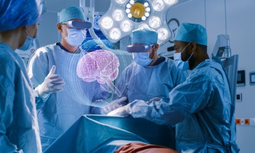
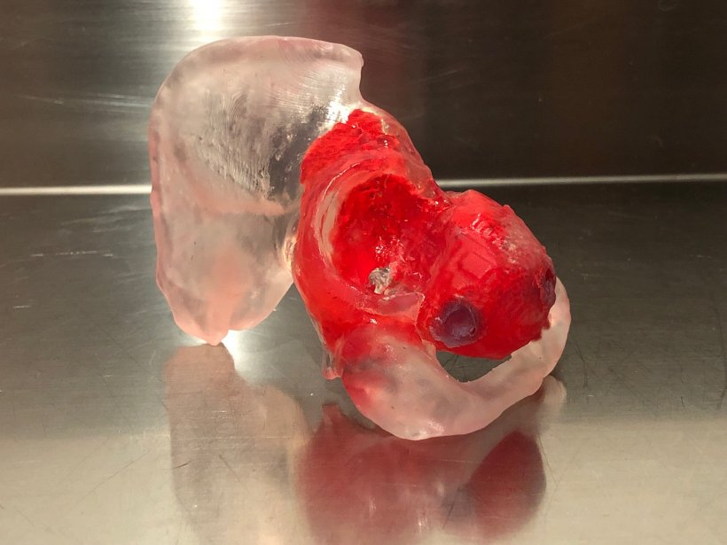
In his lecture, Brantner introduced three main concepts with which 3D printing can help the surgeon: First, anatomical models help the surgeon to visualize complex or invisible structures and help summarize the abundance of image data. They can help the surgeon to improve anatomical understanding in complex situations and provide data for virtual and augmented reality systems. Second, virtual surgery uses imaging data to create patient-specific jigs and to prepare guides as well as patient-specific implants. Third, training models and simulators: Provide tools to educate residents to become more proficient in common procedures while helping experienced surgeons to simulate high-risk interventions.
In an outlook on the future of radiology, Brantner predicted that radiologists “will be faced with a very different way of working.” He said that radiology was perfectly positioned to incorporate 3D printing into clinical routines due to its involvement with nearly all clinical disciplines, “3D printing can add more value to the images that we create.”
Source: Philipp Brantner/3D-Printing Lab at the University Hospital Basel
01.04.2019