News • 3D imaging
New portable gamma ray camera to speed up cancer diagnosis
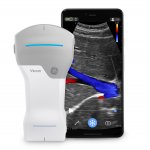
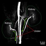
Scientists have designed a portable 3D imaging device which will improve the treatment and diagnosis of cancer. Current handheld gamma imaging tools are small and easy to use, but are limited to providing 2D information, giving doctors and surgeons only part of the overall picture. Much larger systems are able to give three-dimensional images, however, they are bulky and complex – often…