News • PET imaging
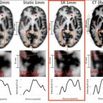
Super-resolution enables detailed brain imaging
A new imaging technique has the potential to detect neurological disorders at their earliest stages, enabling physicians to diagnose and treat patients more quickly.


A new imaging technique has the potential to detect neurological disorders at their earliest stages, enabling physicians to diagnose and treat patients more quickly.
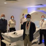
A cutting-edge imaging technique that creates 4D flow images of the heart has been carried out for the first time in the UK at the Norfolk and Norwich University Hospital (NNUH).
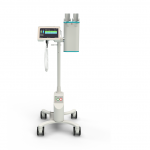
‘Accutron CT-D Vision is the latest evolution of Medtron`s flagship CT injector. Designed to enhance operability, the updated user interface is displayed on larger touch screens and provides a simplified programming and more comprehensive follow-up of each injection step,’ the company reports. ‘The new IDS (Injection Data Sharing) option enables injection data to be shared through RIS/PACS…

In the emerging era of personalized medicine, risk-based breast cancer screening protocols may be better than the one-size-fits all approach. It’s time to consider adopting them, Jack Cuzick, Ph.D., said in the President’s Address of the 2021 Society of Breast Imaging/American College of Radiology Breast Imaging Symposium.
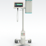
With the launch of the Accutron CT-D Vision, MEDTRON AG once again demonstrates their status as a partner to radiologists. Focusing on the operator's needs, the latest evolution of the Accutron CT-D enhances the usability of its double head CT injector and optimizes its integration into the radiology environment.

SmartXR is designed to make users' work easier and to support them in image acquisition, for example in aligning the DR detector, positioning the patient or setting optimal acquisition parameters. The intelligent assistance systems thus aim to optimize operative and clinical performance, for example. The new robust Dura XD detectors are particularly characterized by their long battery life, and…

University of Washington researchers have discovered that AI models—like humans—have a tendency to look for shortcuts. In the case of AI-assisted disease detection, these shortcuts could lead to diagnostic errors if deployed in clinical settings.

Three leading AI scale-ups - Aidence, ScreenPoint Medical and Thirona - have launched the informative video series “Opening the black box of AI in medical imaging”.
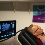
The EU-funded consortium Laser and Ultrasound Co-analyzer for Thyroid Nodules (LUCA) has developed a non-invasive, low-cost device that brings a new solution for thyroid cancer screening.

Guerbet, a global leader in medical imaging, has announced the election of Marc Massiot as director for a six-year term. This nomination was approved at Guerbet's Annual General Shareholders' Meeting on May 28.

One of the negative impacts of the coronavirus pandemic has been the suspension or postponement of many cancer screening services. Each year, these services help prevent and detect the presence of cancer at an early stage; timely diagnosis and care are crucial in preventing the spread of cancer. The National Screening Observatory has confirmed this, reporting alarming statistics: during the first…
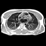
A new project for radiation exposure reduction aims to improve justification of computed tomography (CT) in Europe through co-ordinated action. For this, the European Society of Radiology has been awarded the European Commission Tender ‘European co-ordinated action on improving justification of computed tomography’ (acronym: EU-JUST-CT). The project started on 7 April 2021 and will last until…

Carestream Health is transforming and accelerating the way it develops and delivers AI applications for medical imaging that help improve patient care. The state-of-the-art initiative is based on Hewlett Packard Enterprise’s (HPE) GreenLake for Machine Learning Operations (ML Ops). The machine-learning-optimized cloud service infrastructure makes it easier and faster to get started with ML/AI…
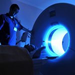
Currently, two opposing trends can be observed in MRI: on the one hand 1.5T scanners are increasingly replaced by 3T scanners for standard clinical MRI applications. On the other hand scanners with lower field strengths have become commercially available in the past years. A session at ECR 2021 took a closer look at low-field (0.5T) and ultra-low-field (0.05T) MRI scanners.

Low-field MRI is the flavour of the season after several 1.5T models were launched. The major advantages of this technology are a relatively low purchasing price and operating costs and few artefacts while the image quality is comparable to a 1.5T scanner. Patient comfort is another benefit of low-field scanners. This advantage, however, tends to get short shrift despite the fact that it might…

Carestream Health has released Smart Noise Cancellation (SNC), a groundbreaking artificial intelligence (AI)-based technology that greatly improves image quality — producing images that are significantly clearer than with standard processing.
A new study from the University of Surrey and University College London has revealed that treatment for heart attacks could be improved thanks to a novel method of evaluating heart function using contrast-based MRI scans. According to the British Heart Foundation, heart and circulatory diseases cause more than a quarter (27 per cent) of all deaths in the UK, which equates to more than 160,000…
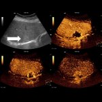
The American Institute of Ultrasound in Medicine (AIUM) and the American Society of Echocardiography (ASE) have joined the International Contrast Ultrasound Society (ICUS) in recognizing the relatively low risk and important clinical benefits of ultrasound contrast agents (UCAs), which are used routinely around the world to help detect heart disease, stratify the risk of heart attack or stroke,…
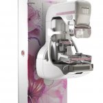
Fujifilm Europe GmbH is launching the new “Harmony” version of its Amulet Innovality mammography system. The “Harmony” version brings together improved diagnostic performance with new design themes to embellish and illuminate your mammography suite, creating an environment to put your patients at ease.
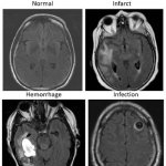
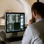
An artificial intelligence (AI)-driven system that automatically combs through brain MRIs for abnormalities could speed care to those who need it most, according to a new study. “There are an increasing number of MRIs that are performed, not only in the hospital but also for outpatients, so there is a real need to improve radiology workflow,” said study co-lead author Romane Gauriau, PhD,…
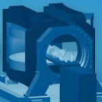
Researchers at the Helmholtz-Zentrum Dresden-Rossendorf (HZDR) want to build the world’s first prototype that tracks moving tumors with magnetic resonance imaging (MRI) in real time during proton therapy. They are combining a rotating open MRI device, designed for the LINAC-MR system from Alberta Health Services, with an actively scanned clinical-akin proton beam at OncoRay, the Dresden-based…
In an industry where every second and every click counts, workflow inefficiencies consume as much as a third of the MRI procedure time. This is a key area of focus where technology advances can radically change what is possible with an MRI exam. Given declining reimbursements, fewer skilled resources, and the system-wide burden of chronic diseases, maximizing productivity is a strategic…

Over the past few years, modern appointment management has found its way into German medical practices. Nevertheless, there is significant room for improvement as some doctors can still be contacted only by phone and only during office hours.
The pandemic has presented healthcare systems with new challenges, resulting in backlogs of routine screenings and delayed procedures which threaten the health and wellbeing of patients, as well as the ability of facilities to serve their communities. In order to address these widespread issues, we need to ensure that healthcare professionals are able to operate with precision, confidence and…

Dunlee announces that it has successfully installed its first CT replacement tubes with liquid metal bearing (LMB): the new DA200P40+LMB tube with Dunlee CoolGlide technology. Prior to this first installations, the DA200P40+LMB tube with Dunlee CoolGlide technology was rigorously tested at both Dunlee's facility and on independent external gantries to confirm that it will perform reliably in both…