News • Potential of nigrosome imaging
Parkinson's: Misconception of 'swallow-tail'-sign rectified
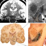
A new understanding of the 'swallow-tail', an anatomical landmark in the brain, could pave the way for earlier detection of Parkinson's disease.

A new understanding of the 'swallow-tail', an anatomical landmark in the brain, could pave the way for earlier detection of Parkinson's disease.

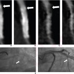
The combination of deep-learning reconstruction and a subtraction technique yields promising diagnostic performance for the detection of in-stent restenosis by coronary CTA.

The portfolio Philips presented at ECR 2022 revealed that the company not only advanced their products, but also listened to medical professionals and patients – and took their feedback to heart.

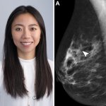
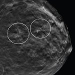
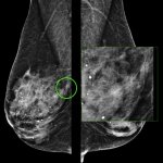
Patients with multiple architectural distortion (AD) identified on digital breast tomosynthesis may benefit from biopsy of all areas, given the variation of pathologic diagnoses across individuals.

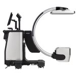
At the European Congress of Radiology (ECR) in Vienna, Fujifilm Europe showcased a new flexible, hybrid C-arm and portable x-ray machine, called FDR Cross.

Researchers are seeking alternatives to gadolinium-based contrast agents (GBCAs) to help raise levels of patient safety. An open hybrid session at ECR in Vienna heard how research across several centres has been examining the options of new approaches to reduce reliance on GBCAs.

A Europe-wide project aims to improve medical radiation protection for patients and staff by better understanding and evaluating the health effects of exposure to low-dose ionising radiation resulting from diagnostic and therapeutic procedures.

Leading radiologists have debated whether working from home is a blessing or a curse amid the evolving impact of teleradiology. In a hybrid ECR session examining working from home in radiology, speakers discussed their own experiences.

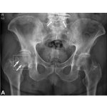
Fujifilm partners with Gleamer to integrate an AI software called BoneView into its X-ray imaging systems to assist radiologists and emergency clinicians in the diagnosis of skeletal fractures.

Dark-field radiography uses a part of X-rays that has hardly been considered so far to expand the spectrum of diagnostic imaging. At the ECR Overture, Theresa Urban presented the new method.

Contrast-enhanced mammography (CEM) has gone a long way since it was first approved by the Food and Drug Administration (FDA) in 2011. An Italian expert explained how useful the modality has become in breast imaging in a dedicated course at ECR 2022.

Building artificial intelligence (AI) tools that clinicians and patients can trust, and easily use and understand, are core to the technology being successfully deployed in healthcare settings.

AI-based diagnosis is undoubtedly one of the most promising subjects when we talk about the future of radiology. Now, a couple of new studies indicate that most radiologists are open to using the technology and this for good reasons.

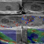
Multiparametric ultrasound (MPUS) has proven its value in the abdomen – now, the technique is increasingly moving towards peripheral areas such as breast and testis imaging, experts showed in a dedicated session at ECR 2022.

Rapid technical advances in ultrasound and increasingly tight healthcare budgets call for a change in ultrasound practice, experts explained in a session at the ECR in Vienna. Two experts presented possible models for the next two decades.

After two online meetings and a teaser last March, the European Congress of Radiology (ECR) is coming home to Austria in July 13-17. We spoke with congress president Regina Beets-Tan about how radiologists must build bridges to go forward.

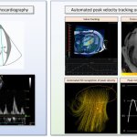
Researchers have developed cutting-edge imaging technology to help doctors better diagnose and monitor patients with heart failure. The state-of-the-art technology uses magnetic resonance imaging (MRI) to create detailed 4D flow images of the heart.

Using functional MRI with inhaled xenon gas, researchers have identified that long COVID symptoms are related to microscopic abnormalities that affect how oxygen is exchanged from the lungs to the red blood cells.

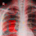
Physicians use AI-powered technology for faster and earlier detection of diseases. At ECR Overture, Dr Steven Schalekamp, PhD, discussed the application of AI for chest radiography in paediatrics.

The classification of brain tumors—and thus the choice of optimal treatment options—can become more accurate and precise through the use of artificial intelligence in combination with physiological imaging.

Incorporating AI support into clinical practice can reduce repetitive tasks, saving approximately one hour of chest CT interpretation time in a radiologist’s typical workday.

Medical imaging and radiotherapy treatment devices are essential lifesaving technologies. In an era when hospital information systems are increasingly being attacked by cybercriminals, just how protected and safe is this equipment?

Researchers visualize brain inflammation using diffusion-weighted MRI. This detailed "X-ray" of inflammation cannot be obtained with conventional MRI, but requires data acquisition sequences and special mathematical models.

Cerebral white matter hyperintensities (WMH) can have a devastating impact on a patient’s life. Radiologists must learn to recognise these white spots in the brain and conventional imaging may just reveal the tip of the iceberg.

Advances in image post-processing and contrast-enhanced techniques have widened the scope of possibilities for MRI and ultrasound vascular imaging, experts showed in a dedicated course at the ECR Overture.