Non invasive but effective
A Canadian study published in the New England Journal of Medicine concludes that chest pain in patients with heart disease could be treated as effective with medication over time as with an expensive angioplasty.


A Canadian study published in the New England Journal of Medicine concludes that chest pain in patients with heart disease could be treated as effective with medication over time as with an expensive angioplasty.

The probability of woman of suffering from cardiovascular diseases is often underestimated. A recent study evaluated the risk of nearly 9.000 women in the U.S. screened for heart-health risk: one in five women had a higher risk than measured by a frequently used predictor.
A pressure sensor that is implanted into the heart works with an electronic monitoring system that wirelessly measures patient's pulmonary artery pressure. It allows physicians to track the patient's pulmonary artery pressure while they remain at home

Hospital admission might probably determine the severity of heart failure. By analysing data from 260 hospitals across the United States researchers created a model to reduce in-hospital mortality and more quickly identify triage methods and treatment decisions.

Compared to 1997 people aged 60 and over receive a lot more from their physicians: Not more attention but more drugs or other medical aids. The average number of prescriptions for elder people doubled from 1997 to 2007, a report from The NHS Information Centre reveals.
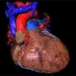
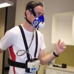
Treadmill exercise testing is a common tool to detect of cardiovascular diseases. But clear images of the working heart are hard to obtain. Now researchers designed MRI equipment to provide high-resolution images of the heart at critical stages.
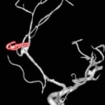
Tiny lipid-shelled microbubbles injected in vessels may serve as an new ultrasound contrast agent to evaluate microvascular blood flow. By this microbubbles US-researcher devised a ultrasound imaging technique that can spot early signs of PAD.

This May the Brussels-based Crossroads Institute for Cardiac and Vascular Medical Education launched two new educational courses on the prevention of amputation (peripheral vascular disease) and on improving the treatment of women with cardiovascular disease.
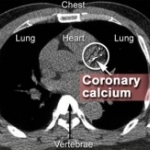
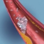
Scanning the heart's arteries for calcium deposits might be one of the best ways to predict the overall risk of death for adults with cardiac trouble, a new study suggests. This might also help end the controversial discussions about calcium scans.
Technologically advanced, cost-effective cardiovascular monitoring systems are increasingly in demand in Europe. New analysis from Frost & Sullivan point out that the market will grow from $350.0 million in 2007 and to $491.3 million in 2014.
Even in 2004, the medical costs for the care of stroke patients in Germany came to 7.1 billion euro. The neurologist Tobias Neumann-Haefelin of the Johann Wolfgang Goethe University in Frankfurt and his colleagues have calculated the projected number of strokes in the German federal state of Hesse for the year 2050.
The ankle brachial index, a ratio of blood pressure measurements used to indicate the risk of peripheral artery disease and atherosclerosis, may be useful to improve the accuracy of cardiovascular risk prediction, according to a meta-analysis of previous studies.
The Cardiomobile developed by the Institute of Health and Biomedical Innovation at Queensland University, Australia, contains a Mini ECG and a GPS system linked to a mobile phone via Bluetooth. That way heart patients can do rehabilitation exercises any place and any time they want to.
A research team at Cedars-Sinai Medical Center, the University of Texas Medical Branch and RWTH Aachen University in Germany has developed a new classification system devised to guide physicians treating patients with symptomatic myocardial bridging, published in the online edition of Cardiology.
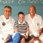
Ten-year-old Tobias smiles brightly at the photographer. And so do the two physicians next to him. They have good reason to be proud as they were the first surgeons worldwide to treat a very serious congenital heart defect in a child by guiding a catheter with the help of a magnetic navigation system into small lung vessels.
For the third in his series of articles for European Hospital, Professor Stefan Schönberg of the Institute of Clinical Radiology and Nuclear Medicine (IKRN), University Hospital Mannheim, Medical Faculty of Mannheim, University of Heidelberg, invited colleagues at the Faculty's Cardiology and Radiology and Nuclear Medicine departments for a round-table discussion on:
Drawing together radiologists from all of Russia is a challenge - even more surprising is meeting the president of the European Congress of Radiology (ECR) and other well-known radiologists from the rest of Europe writes Meike Lerner, of European Hospital, who was at the 2nd National Russian Radiology Congress held in Moscow this May, to report on the hot topics in radiology over the eastern…
The RWTH Aachen University Hospital and the Maastricht University Hospital with their corresponding medical faculties are located centrally in the EU region Meus-Rhine.
According to Prof Clemens von Birgelen, cardiologist on staff at the Thorax Centre of the Medisch Spectrum Twente in Enschede, who quoted from recent studies in his inaugural as a professor at the University of Twente at the beginning of June, this might be the case.
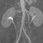
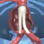
Developments in MRI over the last few years have revolutionized the diagnosis and therapy of cardiovascular diseases. Contrast-enhanced MR angiography has established itself as a non-invasive, standard procedure for the diagnosis of vascular diseases in the thorax, abdomen and periphery.
Cardiovascular diseases kill more than 12 million people worldwide every year and are the cause of death for more than 50% of all Europeans over the age of 65.
In his lecture Focus on new therapy strategies for heart attacks, Prof. Hans Michael Piper, President of this year's Congress of the German Cardiac Society, gave some fascinating insights in to therapeutic measures after heart attacks.
The Department for Paediatric Cardiology at the University Medical Centre, at Johannes Gutenberg University, in Mainz, has extended its range of services for lower impact treatment According to the birth register in Mainz, an annual 1.26% of newborns are diagnosed with congenital heart defects, making these the most common malformation.
The French Health Authorities announced in February that the smoking ban — which began in February 2007 for communal buildings and work places, and was implemented in January 2008 with effect on bars, restaurants and hotels — has produced striking results.
Arrythmogenic remodelling of the left atrium is a common complication of atrial fibrillation, leading to severe haemodynamic disturbances.