Magnetic Field Imaging improves cardiac diagnostics
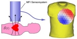
Magnetic Field Imaging (MFI) provides cardiologists with an additional tool to detect arrythmia and irregular cardiac blood flow and thus contributes to a more precise diagnosis. While an ECG acquires electric signals produced by the activity of the cardiac muscle, MFI measures the electrophysical function of the heart by determining the magnetic field during a heartbeat.
“Whereas electric signals are influenced by the differences in electric conductivity of the tissues on their way to the surface of the body, which in some cases they do not even reach, magnetic signals pass practically unperturbed through these regions and thus allow reliable conclusions regarding their source”, explained Torsten Krümmel, founder and managing director of BMDSys, Jena, Germany, who developed the technology.
The system simultaneously records changes of the magnetic field with up to 8,500 frames per second of the region of interest. That means the physician can watch the patient’s heartbeat in slow motion and detect even minor abnormalities. Compared to other modalities such as CT, MRI or ultrasound MFI offers several advantages. First of all, MFI is radiation free and no contrast agents are needed. Moreover, the method supplies functional images of the whole heart instead of showing single slices at a time and offer merely anatomic images.
Currently, a prototype of an MFI system is used for research purposes at the University of Jena, Germany. In collaboration with BMDSys, the researchers have recently developed a new software tool with which provides the physician with the diagnostic results within one minute. Depending on the disease pattern, the software offers several options to display the data, for example in special curves or in 3D.
How does MFI work?
Changes of magnet fields in the human body are one million times weaker than that of the earth. Obviously, measuring those changes requires highly sensitive magnetometers, for example the Superconducting Quantum Interference Device (SQID) which can be cooled down to -269°C, a temperature that allows the detection of human magnetic fields. Single sensor elements amplifiy and digitize the signals with a special low-noise electronic system. A high-end pre-processing system prepares the collected data for analysis. Although the idea behind MFI was conceived about thirty years ago, the problem so far has been the highly complex use of the technology.
BMDSys and Apollo CXS have now successfully developed a user-friendly tool which can be operated by the hospital staff and which supplies standardized results for a quick and safe diagnosis.
09.11.2007
More on the subject: