
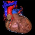
AZ Sint-Jan AV Hospital in Bruges adapts to the new IMPAX Cardiovascular Suite
Among its many specialties, the 909-bed AZ Sint-Jan AV Hospital in Bruges, Belgium, has a high level of expertise in cardiac catheterisations and electrophysiology.


Among its many specialties, the 909-bed AZ Sint-Jan AV Hospital in Bruges, Belgium, has a high level of expertise in cardiac catheterisations and electrophysiology.
Cardiovascular diseases kill more than 12 million people worldwide every year and are the cause of death for more than 50% of all Europeans over the age of 65.

Royal Philips Electronics recently announced it will acquire TOMCAT Systems Ltd., based in Northern Ireland. Terms of this acquisition were not disclosed. TOMCAT offers a software solution to collect and aggregate data relative to the cardiac care of patients, and allows for a comprehensive, patient-centric presentation of this data to care givers such as doctors and nurses.
Today the key results of the Heart Failure Home Care trial, a study designed to assess the impact of a home-based heart failure monitoring system on healthcare costs for heart failure patients will be presented at the American College of Cardiology's 57th Annual Scientific Session (ACC.08).
One of the most painful problems of modern Russia is the high death rate, especially among able-bodied people. In 2005, the average life expectancy for men was 58.8 years.
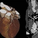
Encouraged by the success of the Dual-Source CT system Somatom Definition with two X-ray tubes that simultaneously generate different energies, Siemens Healthcare has already developed six specific dual energy applications. At the ECR 2008, the company presents four new applications that simplify the diagnosis of diseases of the heart, brain, lungs and extremity joints.

'How do you treat the HIV-positive, diabetic, schizophrenic patient presenting with chest pain? By making the necessary information available for personalised medicine'
Competitive or complementary? By Florian Schwarz BS, Balazs Ruzsics MD PhD and U. Joseph Schoepf MD, of the Radiology and Medicine Departments, at the Medical University of South Carolina, Charleston, USA.

Imaging in Internal Medicine is among the main topics for 114th Congress of the German Society of Internal Medicine (March. Wiesbaden). Specialists in internal medicine, radiologists, and nuclear medicine have developed a programme that will not only provide an overview of the values of modern imaging procedures but also tackle controversial subjects.

As announced today, MEDRAD, an affiliate of Bayer HealthCare and a leading provider of contrast injection systems used to diagnose cardiovascular disease, has entered into a definitive merger agreement with Possis Medical, leading provider of mechanical thrombectomy devices used to treat narrowed or blocked blood vessels. MEDRAD will acquire Possis Medical in a cash tender offer for US-Dollar…
According to a study that was recently published on bmj.com, researchers found evidence that a significant number of patients treated with low doses of aspirin for its antiplatelet effect are actually aspirin resistant. Such a resistance makes patients four times more likely to suffer a life-threatening stroke or heart attack.

A new test, developed at the University of Leeds, searches for a heart-type fatty acid-binding protein (H-FABP) which is released into the circulation following heart injury. The test seems to be more accurate in identifying patients with heart damage at an earlier course of their illness.
During a workshop organized by the European Science Foundation (ESF), researchers discussed further steps in developing computerised “in-silico” models of the heart that simulate the real heart and enable possible drugs and therapies to be tested without risk to people.

According to a recently published study in the Journal of Bone and Mineral Research, a vertebral fracture assessment (VFA) examination can be used to measure abdominal aortic calcification (AAC). The level of AAC can predict the likelihood of myocardial infarction as well as stroke among elderly women, independent of other clinical risk factors.

A research carried out at six hospitals in the West Midlands, UK, is evaluating the use of pulse oximetry as a screening tool for congenital heart disease in newborn babies. Approximately three percent of infant deaths are caused by these significant heart defects and at the moment only less than half of the affected babies are identified by clinical examination, the current screening technique.
Recent studies show that drug-eluting-stents perform superior than bare metal stents in the treatment of high-risk cardiac patients. At ComPaMED 2007, the Germany based company HEMOTEQ, leading designer and manufacturer of ultra-thin coatings for medical devices, presented their solutions for customized coating of stents.
Poster data presented at Scientific Sessions 2007 have demonstrated that the application of clinical practice modifications, combined with advanced electronic technologies, can improve the care of patients at risk for sudden cardiac arrest (SCA).
Just in time for Medica, VISUS has expanded its products portfolio with a PACS solution for cardiology.
USA - A team of researchers at the Rochester Institute of Technology, is working to advance the integration of radio frequency identification technology (RFID) into existing cardiac sensor networks, a new wireless technology for telemedicine delivery, and will also work to enhance the security of the systems used.

Sweden - According Canadian physician Neil Skjodt a simple MP3 recording device gave a superior performance in auscultation of lung and heart sounds.
The new £2 million remote access stereotaxis catheter laboratory at London's Heart Hospital, funded by the British Heart Foundation (BHF), is the first of its kind in the UK and one of only 40 worldwide.
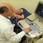
Toshiba's new Artida Ultrasound system is the world's first ultrasound system that can track and display myocardial wall motion three-dimensionally.

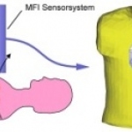
Magnetic Field Imaging (MFI) provides cardiologists with an additional tool to detect arrythmia and irregular cardiac blood flow and thus contributes to a more precise diagnosis. While an ECG acquires electric signals produced by the activity of the cardiac muscle, MFI measures the electrophysical function of the heart by determining the magnetic field during a heartbeat.
Magnetic Field Imaging (MFI) provides cardiologists with an additional tool to detect arrythmia and irregular cardiac blood flow and thus contributes to a more precise diagnosis. While an ECG acquires electric signals produced by the activity of the cardiac muscle, MFI measures the electrophysical function of the heart by determining the magnetic field during a heartbeat.
The Eastern Lithuania Cardiology Project (ELCP) - an integral inter-institutional regional project sponsored by the Lithuanian Government and the European Structural Funds, which began in 2004 - will end this year. In May, those who voted on the Lithuanian EU Support official website (a specially organised event, focusing on all EU-supported projects in all fields) nominated this project as the…