News • Magnetic resonance imaging
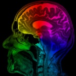
Multicolor MRIs could aid disease detection
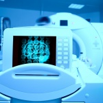
Researchers at Case Western Reserve University School of Medicine have developed a method that could make magnetic resonance imaging—MRI—multicolor.


Researchers at Case Western Reserve University School of Medicine have developed a method that could make magnetic resonance imaging—MRI—multicolor.
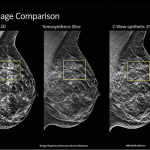
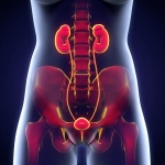
The European Society of Breast Imaging (ESOBI) promotes high quality breast imaging across Europe by developing education and training, encouraging research and promoting guidelines and standards. This year’s meeting (23-24 September) was held in collaboration with the French Society La Société d’Imagerie de la Femme (SIFEM) and drew around 600 radiologists. The event included a two-day…

As the demand for imaging studies booms and digital pathology takes off, it is becoming necessary to look at the interactions between radiology and pathology in telemedicine, an expert explained during the Radiology Triangle Madrid meeting early this year.
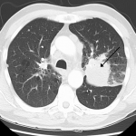
Lung cancer is the cancer that causes the highest mortality worldwide, claiming an estimated 1.7 million patients annually, with 270,000 of those deaths in Europe.
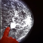
Biannual mammography can help to reduce breast cancer mortality by 40% in women aged 50-69, but the benefits for women under 35 years old are questionable, eminent Spanish radiologists highlighted during a session held by SERAM, their national society of radiology, last November, during the International Radiology Day.
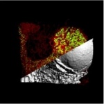
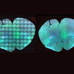
Scientists from the University of Würzburg give fascinating 3D-insights into the bone marrow, and successfully elucidated new details about the process of thrombocyte generation.
Olympus is celebrating art in science with a competition to find the best light microscopy images taken in the EMEA region this year.
The U.S. Food and Drug Administration cleared the first MRI device specifically for neonatal brain and head imaging in neonatal intensive care units.
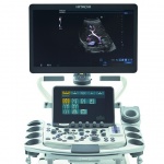
Hitachi introduced the world’s first practical implementation of the CMUT (Capacitive Micro-machined Ultrasound Transducer) silicon wafer technology in 2009. ALOKA ARIETTA 850 exploits the next-generation of CMUT technology, and, combined with eFocusing, a dynamic transmission and reception technology, achieves outstanding clarity of imaging from near to far field. Additionally, this …
Bracco Imaging S.p.A., a company in the diagnostic imaging business, announced today that its contrast agent SonoVue (sulphur hexafluoride microbubbles) is the first ultrasound contrast agent to obtain approval in the European Union for its use in ultrasonography of the urinary tract for the evaluation of suspected or known vesicoureteral reflux in pediatric patients.

Billy Boyle, Founder and CEO of Owlstone Medical, a diagnostics company developing a breathalyzer for disease, is to be awarded the Royal Academy of Engineering’s prestigious Silver Medal. The award recognizes engineer Billy’s work in spearheading the development of the company’s Breath Biopsy platform and driving a vision to save 100,000 lives and $1.5 billion in healthcare costs.

Breast implants may impede an electrocardiogram (ECG) and could result in a false heart attack diagnosis, according to research by the European Heart Rhythm Association (EHRA), a registered branch of the European Society of Cardiology (ESC).
Today, tracking the development of individual cells and spotting the associated factors under the microscope is nothing unusual. However, impairments like shadows or changes in the background complicate the interpretation of data. Now, researchers at the Technical University of Munich (TUM) and the Helmholtz Zentrum München have developed a software that corrects images to make hitherto hidden…

Gadolinium-based contrast agents are an essential component of MRI exams, but are challenged by findings of residual depositions of gadolinium in the body, even though the clinical relevance remains unknown. Three clinicians described how changes to MRI protocols and dose levels for contrast media can optimise the balance between benefit and risk for patients and radiologists.
Current advances such as phase-contrast CT are taking medical imaging further but their use in clinical practice may have to wait up to a decade, prominent physicist predicts.

Positron Emission Tomography (PET) is helping to provide MS experts some insight into what drives progression of the disease. PET also has the potential to quantify the effects of new, targeted therapies on MS patients, according to consultant neurologist Professor Bruno Stankoff.

Ultrasound is often the first line of imaging used in the diagnostic pathway of a patient’s journey into hospital. Additionally, the increased prevalence of chronic conditions and changes in the demographics of the general population has led to an increased demand for ultrasound. Fast-growing advances in technology also shift ultrasound into a more prominent role in patient diagnosis and…
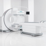
At the 2017 annual meeting of the Society of Nuclear Medicine & Molecular Imaging (SNMMI), June 10-14 at Denver’s Colorado Convention Center, Siemens Healthineers debuts Symbia Intevo Bold, a system that combines the company’s proven single-photon emission computed tomography (SPECT) technologies with new, high-performance CT capabilities to enable a wide range of clinical applications.
Using computed tomography (CT) to evaluate muscle health may help identify optimal treatments for older patients who fall and break their hips, a new study led by radiologists from UC Davis and Wake Forest Baptist medical centers has found.
Toshiba Medical proudly announces its 2nd MRI User Meeting in collaboration with Clinica Creu Blanca in Barcelona on 22 & 23 September 2017 (at Camp Nou FC Barcelona, Spain). At this MRI User Meeting, international experts will share their experiences and clinical solutions for successful diagnostic imaging in Women's and Men's Healthcare.
Anaesthetists working in perioperative medicine have increasingly taken a whole body approach to patient evaluation known as TUBE – Total Ultrasound Body Examination – thanks to the development of point-of-care ultrasound. Dr Christophe Aveline, Consultant Anaesthetist in critical care and surgery at the Sévigné Private hospital in Rennes, is an advocate of TUBE and works closely on its…
Breast cancer continues to be one of the biggest killers of women globally. Worldwide it is estimated that around 522,000 women died from breast cancer in 2012. This is despite the fact that if a cancer is detected at less than 1cm in size with no lymph involvement survival rates at 5 years are comparable with someone who has not had cancer.
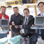
FUJIFILM SonoSite has given two M-Turbo point-of-care ultrasound systems to the non-governmental sea rescue organisation Proactiva Open Arms, based in Badalona, near Barcelona, to support its efforts in rescuing refugees.
Point-of-care (POC) ultrasound is now commonly used in emergency departments throughout the UK. These instruments provide valuable insight for the assessment of both trauma and non-trauma patients, as well as helping to guide procedures. But for many departments, the use of POC ultrasound is limited by a lack of training and poor instrument availability. Professor Bob Jarman, Consultant in…
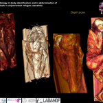
Forensic radiology was, yet again, a central theme at ECR 2017, as Italian radiologists unveiled details of their work in the investigation following the shipwreck in the Mediterranean in April 2015.