Photoacoustic imaging
Promising option for noninvasive monitoring of prostate cancer
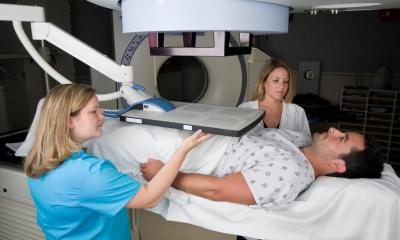
While active surveillance is often recommended for patients with nonaggressive prostate cancer to reduce unnecessary treatment, the challenge for clinicians is to monitor and distinguish early-stage tumors from advanced cancers. A team of scientists led by researchers from Roswell Park Cancer Institute have demonstrated that photoacoustic imaging (PAI) may be an effective tool for more accurately viewing and monitoring prostate cancer.
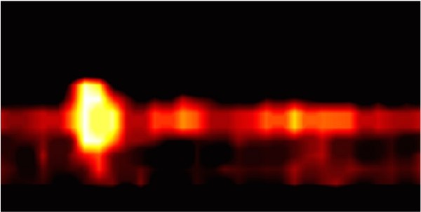
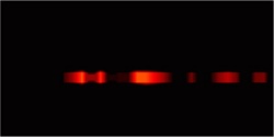
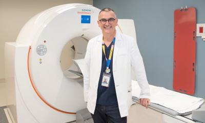
Photoacoustic imaging is an emerging noninvasive imaging modality that has not yet been used in clinical settings. Using photoacoustic imaging, this team of scientists focused a laser light on prostate cells and then “listened” using ultrasound technology to see how a dye attached to a specific prostate cancer marker, PSMA, reacted to the light waves. They chose to study this technology’s use in imaging prostate cancer, as the prostate can be imaged in situ. Photoacoustic imaging of these prostate cells, the researchers found, enabled good discrimination between cells with and without the cancer marker.
“This proof-of-concept study demonstrates that this technology may allow for real-time monitoring of prostate cancer in patients during the course of active surveillance. For patients with more aggressive disease, the technology could offer more precise targeting of biopsies to confirm the need for definitive therapy,” says senior author of the study Kent Nastiuk, PhD, Assistant Professor of Cancer Genetics and Genitourinary Cancers at Roswell Park. “This technology offers the potential to confirm the initial prostate cancer diagnosis, guide biopsies and monitor tumor volume — which is currently not measureable — for improved case management and treatment decision-making.”
Source: Roswell Park Cancer Institute
18.07.2016