Technology
First ultra high frequency ultrasound system for human studies
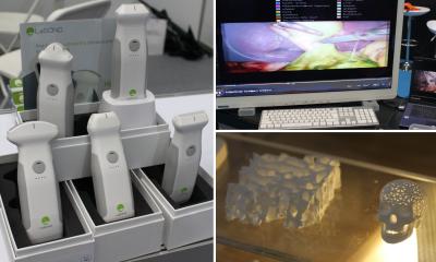
FUJIFILM VisualSonics Inc. announced a significant milestone in its history with the installation of the first Vevo® MD Ultra High Frequency (UHF) clinical ultrasound system at the Second University of Naples in Italy. With multiple successes in preclinical research over the last decade, the Vevo® MD is FUJIFILM VisualSonics’ first foray into the clinical market.
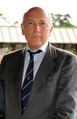
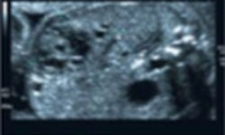
According to Dr. Roberto Grassi, Professor of Radiology at the F. Magrassi and A. Lanzara Department at the Second University of Naples, the institution has used VisualSonics ultra high resolution ultrasound systems for preclinical studies for approximately 10 years. These UHF systems have enabled the research team to study the anatomy of small animals, such as mice and rats, with a resolution down to 30 microns. The addition of Vevo® MD—which has been in use at Second University for about a month now—brings the team’s capabilities to a whole new level and shows great potential for developing novel applications in the clinical sector.
“Preclinical research is a crucial step toward establishing clinical applications, but until the launch of the Vevo MD, there were no clinically-approved ultra high frequency systems for human studies,” said Dr. Grassi. “The Vevo MD is the world’s first CE-marked, ultra high frequency ultrasound imaging system—up to 70 MHz—for clinical use, and will transform our clinical studies, enabling visualization of superficial structures in the human body.”
First commercialized in Europe, the Vevo MD is truly a unique ultrasound system, as it operates at much higher frequencies than any conventional ultrasound system currently available. It allows medical professionals to see what they have never seen before—unparalleled image resolution down to 30 microns. The Vevo MD system is compatible with FUJIFILM VisualSonics UHF series of transducers. This patented transducer technology is capable of operating in a range of frequencies up to 70 MHz, a tremendous increase in resolution compared to conventional ultrasound systems.
The Vevo MD was designed to play a role in a range of clinical application areas including neonatology, vascular, musculoskeletal, dermatology, and other small parts that are within the first 3 cm of the body.
“The beauty of the Vevo MD is that it is non-invasive for our patients and allows us to examine the superficial strata, visualizing structures that we could not see using conventional ultrasound. It has enabled us to determine the finer upper limb damage that occur in arthritis, allowing us to investigate the effectiveness of human drug treatments for these conditions,” said Dr. Grassi. “We are now starting to perform vascular assessment of diabetic patients, and also plan to study dermatological and systemic diseases.”
Source: FUJIFILM VisualSonics
30.06.2016