Product • Light Therapy System
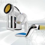
Bioptron Hyperlight Therapy
Bioptron Hyperlight Therapy is a certified medical device that uses a patented form of polarized, polychromatic, non-coherent light. The therapy works by delivering specific light characteristics, which stimulate local blood microcirculation, enhance cellular regenerative process, decrease pain and stimulate immune system. This non-invasive and painless photobiomodulation method is clinically…