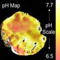
MR images reveal tumour pH
Diseases are often associated with a low tissue pH. Researchers from the UK and Sweden have now developed a MR imaging method to measure the pH in human body using 'baking soda'. The procedure might display the pathological process of inflammation or cancer, as well as the response to the therapy.
A chemical commonly called 'baking soda' which is found naturally in the body could be used to detect cancer with magnetic resonance imaging, revealed a Cambridge study funded by Cancer Research UK and published in Nature.
Magnetic resonance imaging (MRI) is the best method to take a deep look into the body. Traditionally it detects water and fat in the human body. By boosting MRI sensitivity more than 20,000 times - using a scanning technique developed by GE Healthcare - researchers can now image the molecules that e.g. cancer cells use to make energy and to grow – an results in an modified tissue pH.
„A low pH is often associated with the presence of disease“ said Brindle, senior author of the study, to MedicalPhysicsWeb. If one can image tissue pH, this would be a generic method for imaging diseases, e.g. tumours. Although the intracellular pH in tumours is usually normal, outside of the cells it is low.
Working with mice, the team found a new way to measure pH levels using this very sensitive MRI technique with a tagged form of bicarbonate. Bicarbonate, or baking soda, occurs naturally in the body, where it is involved in the acid-alkali balancing system."This technique could be used as a highly-sensitive early warning system for the signs of cancer. Establishing such tools is a major challenge in cancer research“, said Brindle.
"By exploiting the body's natural pH balancing system, we have found a potentially safe way of measuring pH to see what's going on inside patients. MRI can pick up on the abnormal pH levels found in cancer and it is possible that this could be used to pinpoint where the disease is present and when it is responding to treatment."
Using MRI, they looked to see how much of the tagged bicarbonate was converted into carbon dioxide within the tumour. In more acidic tumours, more bicarbonate is converted into carbon dioxide.
First author of the study, Ferdia Gallagher who is a Cancer Research UK and Royal College of Radiologists clinical training fellow, based at the University of Cambridge, said: "Although it's early days, if this technique proves to be safe and effective in cancer patients it has the potential to be a crucial tool in detecting cancer earlier - which is often the key to successful treatment."
Another application for pH imaging is the evaluation of tumour response to chemotherapy. The success otherwise of drug treatment is currently assessed using morphological imaging tools to check for signs of tumour shrinkage. However, it can take many weeks or even months before any noticeable changes in size occur. In addition, some newer cytotoxic drugs do not alter the tumour's bulk volume at all.
"An early read-out of response would be very useful because there are important choices to be made in terms of patients' treatment," Brindle told medicalphysicsweb. Further preclinical tests are planned to see if the hyperpolarized bicarbonate probe could be used in this manner.
Dr Gallagher, who is also a radiologist at Addenbrooke's Hospital in Cambridge, added: "Our technique allows the spatial distribution of pH to be imaged using MRI which is something that has not previously been possible in patients. This new method is important because the chemical we use isn't toxic and is already administered safely to humans."
The pH imaging technique may also have a role in detecting hidden signs of disease that simply can't be seen on morphological imaging, for example, inflammatory processes. Studies are underway to investigate this option.
One possible obstacle to the clinical roll-out of this technique is the rapid speed with which the probe's polarization decays. Once the hyperpolarized bicarbonate is ready for use, it needs to be injected and imaged at once. But Brindle is optimistic that this will be achievable.
"In this particular study, there was a delay of 15 to 20 seconds between getting the hyperpolarized bicarbonate and starting to image. We could work faster than that," he said. "We also expect to get higher levels of polarization in future, which again should buy us more time."
23.07.2008
More on the subject: