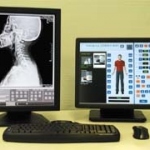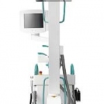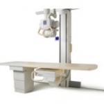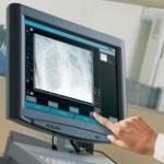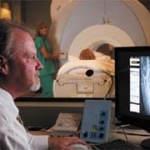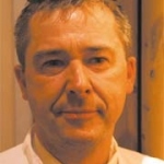
C-Arm technology in daily use
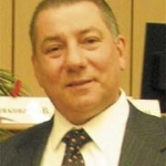
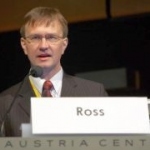
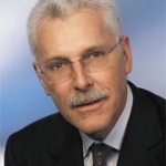
Last year, orthopaedics and sports medicine specialist Dr Rainer Burgkart (left), who is also a musculoskeletal researcher, selected a new C-Arm for use at the Technical University of Munich (Klinikum rechts der Isar). Recently, we asked him for the reasons behind this choice and his subsequent experience with this device.