Every picture tells a story
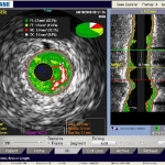
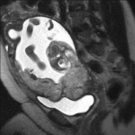
Cardiologists are looking beyond on-screen images of the heart to extract data behind these scans that describe coronary dysfunctions and can reveal hard evidence of the cause of disease reports John Brosky.


Cardiologists are looking beyond on-screen images of the heart to extract data behind these scans that describe coronary dysfunctions and can reveal hard evidence of the cause of disease reports John Brosky.
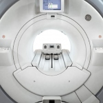
On show in the MR sector at this year’s RSNA was the Optima MR450w wide-bore system manufactured by GE Healthcare. Built on a fully redesigned MR platform, this offers a range of advanced new functions, which, GE reports, makes it ‘a workhorse system for practices of all sizes and specialties’.

A specialised brain area involved in the production of written language was first empirically described by 19th century scientist S. Exner. At that time, the only way to investigate was by post mortem study of patients who had experienced writing problems during their lives. Now, in France, a team of researchers led by Jean François Démonet, has applied state-of-the art technology to study…
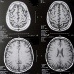
How the brain changes with age is not well-characterized and even less is known about the factors influencing the rate of brain aging. Brain imaging can offer a window into risk assessment into for diseases such as Alzheimer’s disease. A recent study demonstrated that genetic risk is expressed in the brains of even those who are healthy, but carry some risk for AD.
The Alliance for MRI aims to ensure that the threat posed by the EU Physical Agents 2004/40/EC (EMF) to the future of MR is averted and that patients in Europe will not be precluded from state-of-the-art healthcare services. In early 2010 the European Parliament and Council will be sent a proposal from the European Commission to amend Directive 2004/40/EC on electromagnetic fields. This revision…
Using a new noninvasive imaging technique, scientists said they have discovered important, fundamental differences in heart motion by age and gender. Their study - reported in Circulation: Cardiovascular Imaging, a journal of the American Heart Association - is the first to provide gender- and age-specific data on the motions of the normal heart based on a regional analysis of myocardial…
A study presented at the annual meeting of the Radiological Society of North America (RSNA) revealed that MRI is a highly accurate means of identifying placenta accreta, a potentially life-threatening and increasingly common condition that is the leading cause of death for women just before and after giving birth.
At RSNA 2009, GE Healthcare is presenting a big basket filled with trends and innovations for the radiology department:
At this year´s RSNA Siemens Healthcare introduced a new generation of its Tim (Total imaging matrix) technology and its new Dot (Day optimizing throughput) engine. Both technologies are introduced in the new Magneton Aera 1.5 Tesla (T) and the new Magneton Skyra 3T scanners. These two new scanners are the first to incorporate both Tim and Dot technology. The combination of Tim and Dot delivers…
Agfa HealthCare unveiled the latest version of its IMPAX product portfolio at the 2009 meeting of the Radiological Society of North America (RSNA). The improvements to its IMPAX solutions will further increase a radiologist's ability to read more exams, with fewer mouse clicks. Enhancements include new tools supporting multi-planar labeling for volumetric spine studies, new communications tools,…
At RSNA 2009 Visage Imaging will present Visage 7 a single thin client based application that delivers true efficiency for reading everything from plain film to cardiac CT, as well as for state-of-the-art 3D post-processing. Due to its server-based streaming technology all functionality can be accessed efficiently from inside a LAN/WAN and via Internet, on Windows and Mac OS clients. The…
Ziosoft, Inc. a leader in advanced visualization and analysis software for medical imaging will feature its next generation of workflow enhancements and new clinical functionality for the Ziostation system during the 95th Scientific Assembly and Annual Meeting of the Radiological Society of North America (RSNA) at Booth #8353 (North Hall).
Two members of the Heart Center at the University of Leipzig teamed up during Medica for a tour de force presentation on Future Trends in Cardiac Surgery. "The aim of the game is opening the chest through little keyholes to operate in the most minimally invasive way possible and avoid sternotomy," said Prof. Friedrich Mohr, Program Director at the Leipzig Heart Center, who review new surgical…
Philips has developed a proprietary technology for elastography that uses new strain analytics for signals captured on the IU22 volume linear transducer and yields a relative quantification of tissue stiffness, which is presented as statistical charts displayed alongside a colour-coded qualificative image superimposed on the B-mode screen.

Most of the 30 institutes around the world that work with a 7-Tesla MRI scanner do not focus on answering questions about the clinical benefit of this field strength; their efforts revolve around the brain and neurosciences. One exception is the Erwin L Hahn, at the Institute for Magnetic Resonance Imaging, which is based at the Zollverein Coal Mine Industrial Complex, a World Heritage Site in…
The scanner shortage in France is a scandal, says chairman of the French Society of Radiology. New MRIs must be installed in 250 emergency departments
Europe holds a leading position in the research and development of MRI, which has been used for over 25 years, imaging up to 500 million patients without evidence of harm to workers due to EMF exposure. It is also well known that MRI is free from health risks associated with ionising radiation such as X-rays, in many situations the alternative to MRI

A massive scanner for in vivo molecular imaging of the human brain will reveal the metabolic processes of neurological disorders, John Brosky reports
Integrated PET/MRI systems will permit the simultaneous acquisition of molecular, functional and structural parameters. The combined strengths of PET (high sensitivity and specificity, but relatively low spatial resolution) and MRI (high resolution, but low sensitivity) is the most attractive feature of multimodal imaging with hybrid scanners. Their application could substantially contribute to…
If you need to expand your diagnostic imaging services and a fixed installation is too costly but a mobile solution cannot cover your demand, Alliance Medical, Europe's largest provider of out-sourced diagnostic imaging services, reports that it could become your partner. "Did you ever think about a semi-static installation?" the company asks. "The services we can offer by far exceed the…
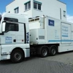
Alliance Medical is Europe´s largest provider of outsourced diagnostic imaging services, with over 15 years of unparalleled operational and service excellence, with offices in the UK, Ireland, Italy, Spain, Poland, Germany and the Netherlands.

The University of Pennsylvania Health System, one of the largest health systems in the US, has awarded a $135 million contract for Integrated Service Management to Siemens Healthcare. Over the next seven years, Siemens will be servicing and ensuring around-the-clock availability of diagnostic systems and bio-medical devices from diverse manufacturers in the customer´s various hospitals.
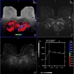
With its new diagnostic reporting software for breast imaging in magnetic resonance imaging (MRI), Siemens Healthcare provides radiologists with new opportunities in Women's Health. The syngo BreVis diagnostic reporting software shows all of a patient's examination results in a single view - for example ultrasound or radiography images next to the images from magnetic resonance imaging -…
After two years of intensive work the results from the German pilot phase of the EuroCMR Register are due to be published in the forthcoming issue of the Journal of the American College of Cardiology*, and also presented and discussed in detail at this year's ECR in Barcelona.
Since its foundation by Michael Friebe PhD in 2003, and on the basis of 10 years of business activity in radiology, Tomovation has become an established provider of high-end services for tomography applications, such as MRI, CT, and PET-CT.