The feasibility of breast MRI at 7 Tesla
Preliminary results
Within the last three decades, contrast-enhanced magnetic resonance breast imaging has gone through substantial developments, evolving to become the imaging modality with the highest sensitivity for breast cancer detection. The indication to perform breast MRI ranges widely, including high-risk patients, dense breast tissue and therapy monitoring under primary systemic therapy.
1.5 Tesla (T) breast MR imaging is considered to be the current clinical standard. After overcoming RF-related limitations of SAR and higher susceptibility effects, recent studies have demonstrated the higher image quality of high field breast MRI in comparison to 1.5 T imaging. The aim of this study was to establish a specific breast examination protocol for a 7 T whole-body MRI system and compare the first ultra high field breast imaging results with standardized clinical 1.5 T examinations.
Methods
10 healthy subjects and 10 patients with breast cancer were enrolled in this study. All subjects were examined on a 7 T whole-body MRI system (Magnetom 7T, Siemens Healthcare, Erlangen, Germany) and a 1.5 T MR system (Magnetom Espree, Siemens Healthcare, Erlangen, Germany) within 48 hours. For imaging of a single breast at 7 T, a unilateral linearly polarized (LP) 10-cm-diameter single-loop transmit / receive coil (Rapid Biomedical, Wurzburg, Germany) was used.
The examination protocol for 7 T imaging included the following sequences 1) T2-weighted TSE sequence; 2) 6 dynamic T1-weighted spoiled gradient-echo sequences (3D FLASH). A gadolinium-based contrast agent (Gadobutrol, Bayer Schering Pharmaceuticals, Germany) was administered with a dose of 0.1 mmol/kg body weight.
For the 1.5 T examinations a standard circularly polarized (CP) bilateral breast coil (Siemens Healthcare, Erlangen, Germany) was used for imaging. The standardized clinical protocol included the same sequence types as for the 7T examination. Subtraction images were generated both at 1.5 T and 7 T. Each sequence at 1.5 T was correlated with the corresponding 7 T data regarding image quality including signal homogeneity, signal penetration depth, and depiction of anatomical structures.
Results
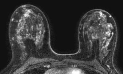
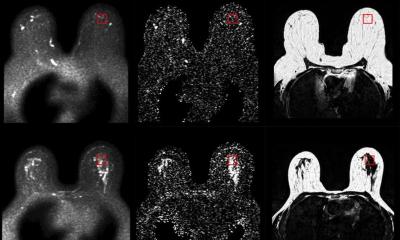
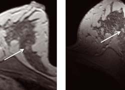
T1-weighted imaging at 7 T could be obtained at a higher spatial resolution with shorter acquisition times, providing higher image accuracy and conclusively a better delineation of small anatomical structures (Fig. 1, 2). After the intravenous administration of gadolinium, the correlation of the contrast enhancement of suspicious lesions revealed a stronger enhancement and superior conspicuity of the tumour lesions at 7 T. Even smaller satellite lesions could be depicted at 7 T that were not demarcated at 1.5 T. T2-weighted imaging could also be obtained with a higher spatial resolution at slightly longer acquisition time. Due to coil limitations, 4 out of the 17 high field MR examinations showed decreased diagnostic value.
Discussion
Our study demonstrates the feasibility and high diagnostic potential of ultra high field breast MR imaging. The higher SNR could be successfully transformed into higher spatial resolution at high temporal resolution, revealing highly detailed anatomical and pathological features. Ultra-high field MRI of the breast remains challenging, as obstacles such as B1 inhomogeneities and physical side effects hamper good diagnostic imaging.
New phased array bilateral transmission/receive coils with efficient use of parallel imaging and inherently higher SNR are expected to circumvent the challenging physical side effects of ultrahigh field strength and to enable high clinical diagnostic imaging in the future.
Authors: Lale Umutlu1,2, Maderwald S1,2, Kraff O1,2, Theysohn J1,2, Kümmel S3,
Hauth EA1,2, Forsting M1,2, Ladd ME1,2, Quick HH1,2, Lauenstein TC1,2
1 Department of Diagnostic and Interventional Radiology and Neuroradiology, University Hospital Essen, Germany
2 Erwin L. Hahn Institute for Magnetic Resonance Imaging, University Duisburg-Essen, Germany
3 Department of Gynaecology and Obstetrics, University Hospital Essen; Germany
07.03.2009