Bordeaux's centre of excellence for sports medicine
Open MRI not only provides good clinical images for musculoskeletal diagnoses, but also makes it easier to accommodate big-bodied sportsmen.

Housed in a smart, modern building in beautiful Bordeaux, the Centre D’Imagerie Osteo-Articulaire, at the Clinique du Sport de Bordeaux-Mérignac, is well known for treating musculoskeletal injuries.
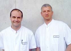
‘We specialise solely in musculoskeletal examinations and interventions,’ confirmed Dr Lionel Pesquer, one of the centre’s eight orthopaedic radiologists, all of whom are qualified in subspecialties such as knee, shoulder wrists or joints ‘Our patients are professionals, semi-professionals and amateurs athletes, such as soccer and rugby players, ballet dancers, skiers.’ Patients travel to the centre from all over Europe.
The centre’s approach is truly holistic. It provides a sports medicine and trauma centre, orthopaedic and sports surgery; a radiology centre that provides traditional and interventional bone and joint radiology, arthrography, ultrasound and MR; a cardiology unit that carries out cardiac stress tests and coronary angiographies for example; a Neurology division providing electro-physiology (EMG) and electro-encephalography (EEG), plus departments focused on Podiatry and Orthotics. In addition, the physiotherapy department, with its team of nine physiotherapists, rehabilitates patients undergoing knee replacement, osteotomy, ACL hamstrings reconstruction or surgery for shoulder instability or rotator cuff pathology.
Equipment in the physiotherapy department includes two isokinetics machines (Cybex norms and Cybex 340), 3 kinetechs, several rehabilitation stations and an area for strength, function and endurance recovery.
The centre also has two X-ray rooms with remote control, and facilities for dynamic X-rays for interventional radiology; a digital, high definition development machine with radio luminescent screen, and ultrasound equipments with high definition probe (until 17 MHZ) - Vein and artery colour Doppler. All the examinations are available on the PACS for internal or external clinicians.
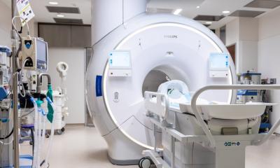
About 16 months ago, the centre opened its MR department, which is now equipped with a 0.35 Tesla open MRI system, the Signa Ovation HD manufactured by GE Healthcare. The system uses a permanent magnet, high definition applications and high-density coils.
‘A kinematic device can be used complementary to the coil so that a scan is made while the patient flexes his knee. For example, it allows better visualisation of the posterior cruciate ligament tears,’ said Dr Pesquer. `The scanner performs also accurate images for the extremities – feet, ankles – which are sometimes difficult to scan. The high resolution images obtained with the MR scanner enable the radiologist to better visualise the lesions and therefore provide enough details to the surgeons to operate.`
‘The performance of the 0.35T scanner is almost equivalent to 1.5T for ligament lesions but remains limited for partial ligament tears and cartilage lesions for which 3T is the gold standard,’ explained Dr Pesquer.
The clinic scans about 23 cases daily. For an open MR 0.35 T, Dr Pesquer explained, the average throughput is about 15 to 17 patients per day. ‘The actual examination takes about 20 minutes, but we make appointments for every 30 minutes, because we have to install the patient, which takes time. So we scan two patients an hour.’
When assessing injuries, the Bordeaux team have found that the openness of the magnets is not only easier for patients but also the user. ‘It resolves the problem of performing musculoskeletal examinations that can be difficult and/or uncomfortable for the patient on cylindrical MR scanners, due to the traditional tunnel constraints,’ Dr Pesquer pointed out.
Laying on the Signa Ovation HD table a patient is surrounded by 210 degrees of open space, which not only makes positioning easier, but helps those who suffer claustrophobia (as well as the elderly and nervous children). However, another vital aspect for the sports medicine centre is that this MRI scanner is far better suited to very large patients, such as rugby players or the bigger athletes.
The centre clearly has a constantly increasing bank of valuable information on injuries and their diagnoses. To ensure such knowledge is shared, it operates a subscription website (www.image-echographie.net, languages: French and English) that is fed new case histories every day to further radiologists’ medical education. The user can simply type in the name of a piece of equipment to find related musculoskeletal cases on every anatomy.
The centre also organises sports medicine radiology workshops, which are held at the premises in Bordeaux. Further details: www.image-echographie.net
20.05.2009


