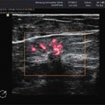
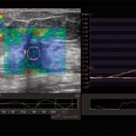
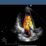
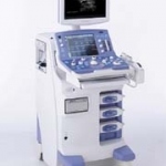
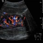
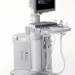
Ultrasound

Photo

A new imaging tool?

Mammography is the common way to detect breast cancer. But it's not perfect: it struggles to image dense glandular tissue or early-stage tumours. Magnetic resonance imaging (MRI) offers best sensivity but it is expensiv and not always specific enough. Now researchers have come up with another option: a scanner that integrates thermoacoustic and photoacoustic tomography to achieve dual-contrast…

Photo

The 2nd National Russian Radiology Congress

Drawing together radiologists from all of Russia is a challenge - even more surprising is meeting the president of the European Congress of Radiology (ECR) and other well-known radiologists from the rest of Europe writes Meike Lerner, of European Hospital, who was at the 2nd National Russian Radiology Congress held in Moscow this May, to report on the hot topics in radiology over the eastern…

Photo

India's world experts on TB

Every three minutes, two people living in India die of tuberculosis. This equates to approximately 370,000 deaths each year, and a staggering economic toll: an estimated US$300 million in direct costs and US$3 billion in indirect costs.

Photo

OSIRIX

Designed by a team of radiologists, the latest release of OsiriX 3.0.1 on the Mac Pro 8-core was demonstrated for the first time at the recent European Congress of Radiology (ECR). OsiriX - a powerful image processing software dedicated to DICOM images (.dcm / DCM extension) produced by imaging equipment (MRI, CT, PET, PET-CT etc.) and confocal microscopy (LSM and BioRAD-PIC format) - a…

Photo

Open High-field MRI

As part of a research and development project, doctors at the University Hospital Magdeburg, Germany, are treating oncology patients with local minimally invasive surgery (MIS) which, for the first time, can be carried out under radiological image control using high-field magnetic resonance imaging (MRI). The system offers excellent image quality under extremely favourable, radiation-free…

Photo

Visualising prostate cancer

Advances in prostate cancer imaging spark hopes for better therapies. Meike Lerner asked Professor Hartmut Huland, Medical Director of Martini-Klinik in Hamburg, and pioneer of the nerve-preserving prostatectomy method, currently the gold standard in prostate cancer therapy, about his technique and whether the optimism regarding imaging is justified or misplaced

Photo

Real-time Tissue Elastography

Today, Real-time Tissue Elastography (HI-RTE) provides a method to determine tissue elasticity of certain organs, such as the prostate, in real-time and to perform precise biopsies for reliable tumour diagnosis during standard examinations.

Photo

Roles in radiology, research, plus a private life?

Apart from spending a year at Johns Hopkins University Hospital, Baltimore, USA, the professor has never worked anywhere other than at the Radiology Department at the University Hospital of Cattinara Hospital, Trieste, of which she is Chairman. Daniela Zimmermann asked her about what women can achieve in this field, as well as the professor's own multifarious roles and research activities.

Photo

Albanian cancer screening-programme for female patients

In the course of the `Interreg III´ Italian-Albanian collaborative project, which is financed by the European Union and the Italian Apulia region, an Albanian screening programme for breast and cervical tumours will be established. The initiative aims at improving the public health system of Albania, regarding the training and the technical equipment needed for such screening projects.

Photo

CAD improves breast cancer diagnosis

A study recently published in the online version of the American Journal of Roentgenology evaluated the clinical value of Computer-Aided Detection (CAD) software in the interpretation of mammograms. In the course of the study, the recall rate, sensitivity, positive predictive value, and cancer detection rate for single reading with CAD, versus double reading without CAD, were investigated.

Photo

New technology enhances X-ray images

An innovative method to produce dark-field X-ray images was recently presented by researchers from the Paul Scherrer Institute (PSI) and the EPFL in Switzerland. Dark-field images offer more details than ordinary X-ray radiographs, allowing to diagnose early stage breast cancer, osteoporosis or Alzheimer's disease.

118 show more articles
Subscribe to Newsletter