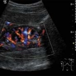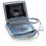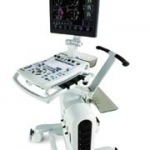
European debut for Acuson S2000
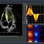
Siemens Healthcare will show its Acuson S2000, the first ultrasound system in the new product series S, at the ESC*. The system platform includes integration of the newest technologies to optimise workflow, e.g. comprehensive software applications such as new software for breast imaging.