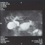
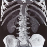
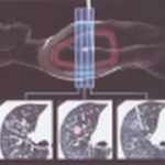
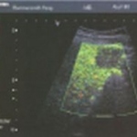
Imaging

Photo

Diagnosis of Cardiac Diseases

For decades, non-invasive cardiac assessment has been at the center of interest. Non-invasive imaging modalities, such as MSCT, MRI, SPECT, PET, and echocardiography, provide valuable cardiac information, and all have been used to measure cardiac morphology, function, perfusion, viability of myocardium, and coronary anatomy for clinical management and research. The last decade brought major…

Photo

Stroke Imaging: Foundations and Strategy

Diagnostic imaging is of particular value in investigating ischaemic cerebrovascular accidents since, due to the rapid progression of cell destruction in the region of the cerebral infarction, the radiologist is faced with urgent questions regarding a possible procedure. These questions may be answered particularly well using computed tomography (CT). The following article provides an overview…

Photo

Dyssynchrony Imaging

Dyssynchrony Imaging (DI), Toshiba's novel Doppler technology, is used as a quantitative clinical tool for rapid assessment of dyssynchrony in patients with suspected myomechanical dysfunction. From the parasternal short axis view, DI is applied by Toshiba's exclusive “angle-correction” technology and in apical views by standard Doppler imaging. Thus, DI permits evaluation of virtually all…

Photo

Stroke Imaging: The Procedure in Practice

In the past, native computed tomography of the neurocranium (CCT) was often the only procedure to diagnose a stroke. Technical limitations of previous generations of scanners made their use impractical for complementary methods of visualising blood vessels or for perfusion imaging. With the introduction of multi-slice spiral CT these technical limitations disappeared and both perfusion CT (PCT)…

Photo

Expanding medical horizons

This was the motto of the ECR 2007 in Vienna, where a group of high-ranking experts discussed diseases of the 21st century; research competition between the US and Europe; the conditions needed to progress leading medical R&D - moderated by Congress President Professor Christian J Herold.

Photo

Turf wars

Nowadays, no radiology conference would be complete without a discussion on turf battles. The rapid development and broad acceptance of cardiac CT has exceeded expectations, and both radiologists and cardiologists are incorporating this technology into their practices. Educational courses on cardiac CT are in high demand and well attended by both radiologists and cardiologists.

Photo

Slovakia

Slovakia was part of the Austrian-Hungarian Empire until 1918, when Czechs and Slovaks were brought together to form the Czechoslovak Republic. After World War II, and up to 1948, the country was still part of Europe, but then fell behind the 'Iron Curtain'. From 1989 it began 'knocking on the EU door' and entry was granted in 2003. Today Slovakia's population is around 5.38 million. To serve…

Photo

Cardiovascular imaging

The US finally did it: radiologists and cardiologists got together to draft guidelines for cardiovascular imaging procedures and define quality standards. However, Europe still awaits a binding consensus to end internal squabbles in hospitals. Meike Lerner, of European Hospital, discussed this situation with Professor Ernst E van der Wall MD, (right) Head of the Department of Cardiology at Leiden…

74 show more articles
Subscribe to Newsletter