Pancreatic Tumours and Echo-Enhanced Ultrasound
Echo-enhanced ultrasound is a newly available imaging modality for use in the differential diagnosis of pancreatic tumours. Ductal carcinomas are often hypovascularised compared with the surrounding tissue. On the other hand, neuroendocrine tumours ar hypervascularised lesions. Tumours associated with pancreatitis have a different vascularisation pattern depending upon their inflammation and necrosis status. Cystadenomas frequently show many vessels along their fibrotic strands. Data from prospective studies have shown that the sensitivity and specificity of echoenhanced sonography in the differentiation of pancreaticmasses are >85% and >90%, respectively, based upon these imaging criteria.
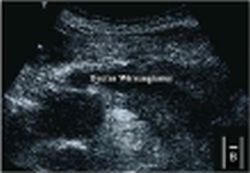
Fig. 1: (b) Poorly vascularised lesion compared to the surrounding tissue using Pulse Subtraction Imaging mode.
This article was first published in the VISIONS, issue 08/2005, a publication of Toshiba Medical Systems
19.07.2007
More on the subject:More on companies: