Tissue Doppler Imaging of the Fetal Heart
In the last 25 years, fetal echocardiography has become established as an integral component of prenatal medicine. Structural heart defects are found in approx. 0.8% of all foetuses. They are consequently comparatively frequent and head the list of isolated fetal organ malformations. Routine screening of the four-chamber view and the outflow tract allows the experienced investigator to detect most congenital heart defects. Considerable improvements in B-image ultrasound and colour Doppler technology in recent years have led to ever more detailed results at earlier and earlier weeks of gestation. Today's high resolution technology allows for cardiac evaluation as early as the 1st trimester.
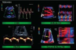
Fig. 1: Examples of use of various TDI techniques on the fetal heart. Top left: C-TDI with velocity curves. Top right: M-mode TDI with sectional
plane through themyocardiumof the right atrium and left ventricle. Bottom left: PW-TDI with Doppler window on the mitral annulus. Bottom right: curvedM-mode DI of the left ventricle in the longitudinal four-chamber view.
plane through themyocardiumof the right atrium and left ventricle. Bottom left: PW-TDI with Doppler window on the mitral annulus. Bottom right: curvedM-mode DI of the left ventricle in the longitudinal four-chamber view.
This article was first published in the VISIONS, issue 9/2006, a publication of Toshiba Medical Systems
12.07.2007
More on the subject:
- cardiovascular diseases (743)
- imaging (1677)
- medical technology (1581)
- obstetrics (150)
- paediatrics (355)
- ultrasound (777)