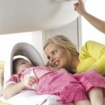
The hospital protocol for dealing with terrorism
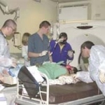
From 2000 to 2007, Israeli hospitals have treated the victims of 148 terrorist attacks.


From 2000 to 2007, Israeli hospitals have treated the victims of 148 terrorist attacks.
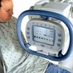
The new Philips 256-slice Brilliance iCT came in to use recently at the University Hospital in Ulm. The system produces quick, high-res scans with 80% less radiation.
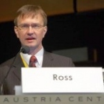
Along with paediatric radiology, interventional radiology will have a high profile at the 89th German Radiology Congress and 5th Joint Congress with the Austrian Radiology Society. Congress presidents Professor Dierk Vorwerk and Professor Richard Fotter outlined what's on the agenda for the expected 6,900 visitors. Training, they pointed out, will aim at those preparing to specialise in…
Designed by a team of radiologists, the latest release of OsiriX 3.0.1 on the Mac Pro 8-core was demonstrated for the first time at the recent European Congress of Radiology (ECR). OsiriX - a powerful image processing software dedicated to DICOM images (.dcm / DCM extension) produced by imaging equipment (MRI, CT, PET, PET-CT etc.) and confocal microscopy (LSM and BioRAD-PIC format) - a…

During ECR 2008, the Canada-based Acceleware Corporation, which develops acceleration solutions for high-performance computing, demonstrated its new AxRecon image reconstruction solution for medical imaging, security, and non-destructive testing.
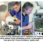
The purchasing and distribution of refurbished equipment was left to specialist retailers for years, until leading manufacturers - for reasons of quality as well as image - established themselves in this business sector. The difference is that these manufacturers not only sell used equipment but also extensively refurbished systems.
The implementation of an integrated HIS/RIS into a single IT platform facilitates data sharing among the more than 1,300 physicians and support staff who work at East Tallinn Central Hospital (ETCH) in Estonia. ETCH, a municipally owned healthcare delivery network, was formed in 2001 with the merger of four hospitals.
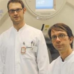
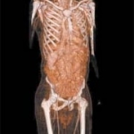
A 320-row CT scanner (Aquilion One, Toshiba Medical Systems Co., Tokyo, Japan) was installed for the first time in Europe, at the Charité University Hospital, Berlin, Germany, in November 2007. Its capability to cover the whole brain in a single rotation means this new type of scanner has the potential to impact strongly on the field of neuro-imaging.
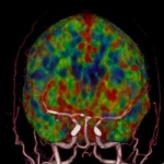
During a Toshiba press conference on Monday at ECR 2008, Prof P. Rogalla Chief Radiologist CT and Prof R. Klingebiehl, Department of Neuroradiology both of the Charité University Hospital, Berlin (Germany), reported about the new diagnostic opportunities the company's latest innovation — the Aquillion ONE - offers.
High definition CT (HDCT) technology developed by GE Healthcare promises to revolutionise image acquisition for CT scanning.
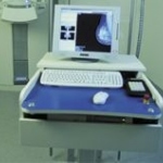
A new volumetric X-ray application, showcased at the European Congress of Radiology in Vienna, Austria, provides physicians with multiple high-resolution slice images of the human anatomy, including the chest, abdomen, extemities and spine.
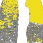
Radiological services and equipment are not yet adapted to obese patients. The accuracy of current MRI, CT and Ultrasound is hindered by subcutaneous and intraabdominal fat. These modalities are crucial in diagnosing pathologies associated with obesity, including heart-related disease. Optimising imaging modalities will be a major challenge for radiology.
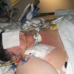
Over the last three decades CT has become a premier diagnostic tool for the evaluation of the acute patient. Over the past ten years in Israel, we have seen an overwhelming increase in the volume of CT examinations in the emergency department (ED).
The Virtual Physiological Human By Hans-Ulrich Kauczor MD PhD, Director and Chairman of Radiology at Heidleburg University Clinic, and radiologists Frederik Giesel MD MBA and Hendrik von Tengg-Kobligk MD of the German Cancer Research Centre in Heidleburg, Germany.
PET/CT imaging exhibits significantly higher sensitivity, specificity and accuracy than conventional imaging when it comes to detecting malignant tumours in children, according to research published in the Journal of Nuclear Medicine (12/07).
Professor Rémy-Jardin MD PhD heads the Department of Radiology and is Chairman of the Department of Thoracic Imaging at the Calmette Hospital, University Centre of Lille. She is also Professor of Radiology in Lille University's Medical Faculty.
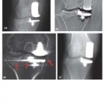
Dr Hiroyasu Yano reports on the effective use of tomosynthesis in orthopaedic surgery.
The healthcare system is in a phase of transition - from planned economy to free market economy. Competition is becoming a challenge. Only entrepreneurs and enterprises that develop creative strategies will stay on top - or make it to the top.
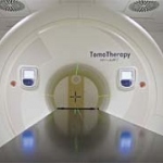
By Rudolf Schwarz and Andreas Krüll, of the Section of Radiation Oncology Department, Ambulanzzentrum GmbH of the University Medical Center Hamburg-Eppendorf
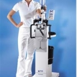
ulrich medical has added a number of new products to the firm's wide range of injectors and accessories for computer and magnetic resonance tomography.
Agfa Healthcare's new IMPAX solution suites offer PACS and RIS to cover hospital data handling and cardiovascular, cardiology, orthopaedics, mammography and radiology data.
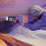
As part of a research and development project, doctors at the University Hospital Magdeburg, Germany, are treating oncology patients with local minimally invasive surgery (MIS) which, for the first time, can be carried out under radiological image control using high-field magnetic resonance imaging (MRI). The system offers excellent image quality under extremely favourable, radiation-free…
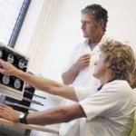
Imaging in Internal Medicine is among the main topics for 114th Congress of the German Society of Internal Medicine (March. Wiesbaden). Specialists in internal medicine, radiologists, and nuclear medicine have developed a programme that will not only provide an overview of the values of modern imaging procedures but also tackle controversial subjects.
Apart from spending a year at Johns Hopkins University Hospital, Baltimore, USA, the professor has never worked anywhere other than at the Radiology Department at the University Hospital of Cattinara Hospital, Trieste, of which she is Chairman. Daniela Zimmermann asked her about what women can achieve in this field, as well as the professor's own multifarious roles and research activities.
US launches a national initiative By Cynthia E Keen