Radiation Treatment Planning
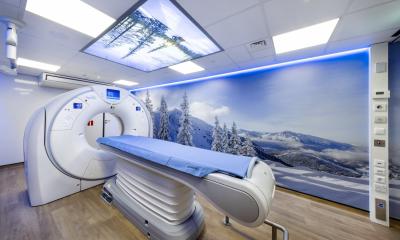
Radiotherapy treatment planning relies on transversal CT images. They form a basis for treatment planning, dose calculation and increasingly the plan localization for external radiation therapy, called CT simulation. A dedicated CT scanner for radiotherapy plan simulation is one of the most essential pieces of equipment in a modern radiotherapy department. In contrast to external beam radiotherapy, treatment planning in brachytherapy has mainly been based on conventional radiographs or fluoroscopy. Brachytherapy is a form of radiotherapy where a radioactive isotope is introduced into the patient's cavities (intracavitary or intraluminal) with special applicators or into the tissue (interstitial) with needles. With the increasing number of CT and MRI scanners in hospitals, 3D image-based planning is now being introduced also to brachytherapy. Because of the 2D history, international dose definitions in brachytherapy are prescribed to points rather than volumes. Recently the recommendations for 3D based treatment planning have been published by Gynaecological (GYN) GEC-ESTRO working group (see literature).

This article was first published in the VISIONS, issue 11/2007, a publication of Toshiba Medical Systems
29.08.2007


