Virtual anatomy
A new way of teaching and learning
A comprehensive knowledge of the topographic relationships of anatomical structures is a must for all doctors.
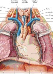
Topographical knowledge used to be taught mainly through the preparation of human bodies and was then put into practice and intensified during surgery.
However, in modern medicine non-invasive imaging procedures, such as ultrasound or split-imaging procedures, e.g. computed tomography (CT) and magnetic resonance imaging (MRI), are now also of decisive importance for diagnosis and therapy planning in non-surgical medical disciplines. To meet this increased demand, even at the training stage, imaging procedures will play a more significant role in the way medicine is taught.
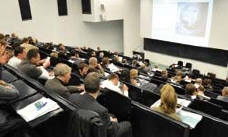
Recently, medical students at the Medical Faculty of University of Heidelberg have been able to learn about radiological split-image procedures right from the beginning of their studies, alongside the more classical learning about anatomy using human bodies. The Institute for Anatomy and Cell Biology recently began to offer a practical course in abdominal ultrasound, along with a ‘Virtual Anatomy’ seminar, in intensive co-operation with the Radiology Department of the German Cancer Research Centre.
The Virtual Anatomy seminar* aims to train students in their ability to interpret and process CT and MRI images at the computer. As the preparation course and the seminar are run at the same time the students can put the different imaging procedures in direct relation to one another. In addition, the use of powerful computers facilitates visual representations known as virtual preparation with the support of advanced medical imaging software (3mensio, developed by the software development company 3mensio Medical Imaging BV, in The Netherlands) (b). Besides this virtual approach 3D print models (supported by VitalRecon) out of digital CT data will enhance the 3D learning in especially in challenging anatomical structures (c).
Both seminars are run like ‘hands-on workshops’ offering practical exercises under the supervision of experienced tutors and lecturers. Upon the conclusion of the very successful trial phase both seminars are now to become integral parts of the core curriculum.
* Initiated and developed by Sara Doll and Frederik Giesel MD, of the German Cancer Research Centre, Heidelberg.
Kindly supported by VitalReconTM and 3mensio Medical Imaging BV.
By Frederik Giesel MD
30.10.2007