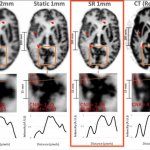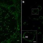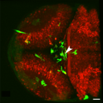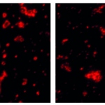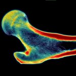
News • AI-assisted microstructure assessment
Super-resolution imaging helps predict bone fracture risk
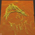
A new AI tool creates super-resolution images showing the inner structures of bones in great detail. This can be used to better determine risk for fracture in elderly patients with osteoporosis.