Article • MRI & Liver
MRI leading the way in metabolic disease imaging
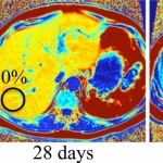
New MRI techniques are set to offer advances in the earlier detection of liver disease in patients. Radiologists are harnessing the potential of highly-targeted MRI, whilst exploring the imaging modality as a means of delivering non-invasive biomarkers, reducing the need for biopsy to measure treatment response.