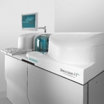
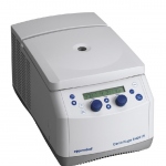
Medical technology

Photo

Turning data into knowledge

What connects these: dementia research; the development of a procedure for fast, cost efficient gene sequencing; protein research; modern IT infrastructures, and the determination of reimbursement structures for medical procedures? Answer: Two extremely small common denominators: Bits and bytes. This means data and, increasingly, mass data, which now forms the foundation of medicine. The question…

Photo

Hybrid operating theatres

In technical terms ‘hybrid’ is a system that connects two technologies so they may benefit from each other. This also applies to the newest generation of operating theatre*: hybrid OTs combine diagnostic and surgical facilities that are usually found in separate locations. Thus procedures can be carried out in less time and involve less discomfort and risk for the patient.

Photo

RSNA 2010

A double-edged sword would be a good analogy for diagnostic imaging in 2010. New ways to utilise imaging technologies are being developed, imaging equipment is doing more, faster than ever, and image processing software is increasingly innovative. Today's radiology exams are ‘slicing’ through the body to reveal anatomy with increasing clarity for better diagnoses and therapeutic treatment…

Photo

MPR 3-D echocardiography

A cardiologist at a UK hospital has become the first in the world to develop a technique to ‘slice’ 3-D images of the heart into intricate sections using computer software. The method, devised by consultant congenital cardiologist Dr Joseph Vettukattil at Southampton General Hospital, is known as multiplane review (MPR) 3-D echocardiography. This allows cardiologists to identify heart defects…

Photo

Real-time tissue elastography as a complementary procedure

Tissue hardness provides radiologists and gynaecologists with significant information to help distinguish between benign and malignant tumours. Tumour tissue is harder and less malleable than normal glandular and fatty tissues. Therefore, the classification of tissue hardness determines whether a biopsy is necessary. For breast diagnoses, real-time tissue elastography, along with conventional…

Photo

The Virtual Autopsy Table

This is imaging with a ‘wow’-effect: The Swedish Centre for Medical Image Science & Visualisation (CMIV) in cooperation with the Norrkoping Visualisation Centre has developed a ‘Virtual Autopsy Table’ that allows a unique look inside the human body and takes interaction with volumetric medical data to a new level.

Photo

EASD 2010 review

7,000 people from 120 countries met in Stockholm this September to hear international experts discuss the progress, solutions and challenges of one of our greatest healthcare burdens. Prevention, self-monitoring, surgery, guidelines, economic problems, drug-safety, and co-morbidities – these are just a few of the problems associated with the care of about 55 million diabetics in Europe.

Photo

Where are we - and what lies ahead?

In a European Hospital interview Professor Herman Requardt, Head of Corporate Technology and CEO of Siemens Healthcare Sector, offered his views on current and future healthcare manufacturing needs as well as the market challenge arising from the dynamic economic ascent of other nations.

Photo

New Partnership

EIZO GmbH, Display Technologies and Ondal Industrietechnik GmbH announce their partnership to provide monitor carrying systems, monitors and accessories as system solutions. These are suitable to use in interventional radiology as well as in standard and hybrid operating rooms.

Photo

Article • MRI

9-, 10- or even 11-Tesla – is more always better?

Advancing magnetic resonance imaging (MRI) up the Tesla scale may sound good, but will it produce the results and patient safety radiologists actually desire? Faced with the question: ‘How many Tesla should it be?’, Professor Siegfried Trattnig MD, head of the Centre of Excellence in high-field MRI at the University Clinic for Radiodiagnostics, Medical University of Vienna, and Austria’s…

Photo

The potentiated value of MR-PET imaging

Even more precise diagnoses, even better process controls -- the future of MR-PET technology has dawned. The first commercial, full-body hybrid scanners are either waiting in the wings or already installed. But what does the introduction of the MR-PET really mean for clinical practice? Professor Heinz-Peter Schlemmer MD, Head of the Radiology Department at the German Cancer Research Centre in…

The MIRACLE begins

Detection of circulating and disseminated tumour cells in blood is a promising method to diagnose cancer dissemination, or to follow up cancer patients during therapy. Today’s methods and involve time-consuming (more than a day) sample processing and cell isolation steps -- all labour intensive and expensive. A lab-on-chip that could integrate those processing steps would enable faster,…

Photo

Microchip separates and extracts tumour cells in the blood

Ikerlan-IK4 and Mondragón Unibertsitatea are taking part in a project initiated by the Spanish National Research Council, CSIC, and which has developed a microchip capable of separating and extracting tumour cells in the blood stream by means of ultrasonic waves. The Foundation General Hospital of the University of Elche, together with researcher Alfredo Carrato, has also collaborated on this…

Photo

Cardiovascular ultrasound

Developed in partnership with international cardiologists, Medison reports that its EKO7 Cardiovascular Ultrasound system is a dedicated Cardiovascular Ultrasound system ‘…with 2-D Image Quality that you would expect from a premium class ultrasound system. As a matter of fact, the same applies to the colour Doppler, Pulsed Wave Doppler and CWD. So the EKO7 has excellent image quality, a…

Photo

Supporting role in the OR

Whenever a clinic sets up a new operating room or intensive care unit, the equipment should be able to meet all requirements for the next 20 years. At the same time, however, tight budgets increasingly restrict many hospitals’ freedom to act.

592 show more articles
Subscribe to Newsletter