The Nanomaxx System
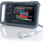
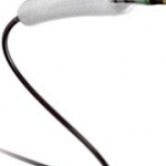
‘Supremely portable and incredibly tough, the NanoMaxx ultrasound system combines Best in Class performance with affordability and simplicity,’ the Erlangen-based manufacturer SonoSite reports.


‘Supremely portable and incredibly tough, the NanoMaxx ultrasound system combines Best in Class performance with affordability and simplicity,’ the Erlangen-based manufacturer SonoSite reports.
The heart of cardiac radiology set the rhythm for this October's 10th Annual Meeting of the European Society of Cardiac Radiology (ESCR) in Leipzig, when state-of-the art technology and progress in cardiac imaging were introduced alongside an educational programme that catered for experienced as well as novice cardiac radiologists
In the last fifteen years, myocardial perfusion SPECT imaging (MPI) has been performed most commonly by dual-head conventional scintillation cameras with parallel-hole collimators, configured in a 90ºdetector geometry and image reconstruction based on standard filtered back projection (FBP) algorithms. Such arrangement, although clinically well established suffers from important limitations…
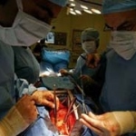
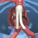
Today, transcatheter aortic valve implantation (TAVI) represents an effective therapeutic alternative to conventional aortic valve replacement for patients who are at high risk or with contraindications to surgery, and the combination of the transfemoral and transapical approaches further increases the number of patients who can be treated.
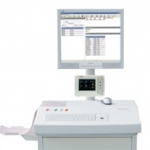
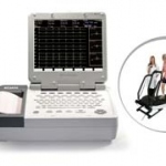
CS-200: SCHILLER´s complete Diagnostic Solution has been redesigned, offering now even more added value. Discreet but important external modifications encompass a large, swivel-mounted 19`` monitor, as well as an ergonomic design. A new, simplified user-interface as well as various new holders and mounting kits for external devices such as gel bottles, bar code scanners, spirometry sensors and…

Since 2007, when a special public information website was set up by the Heart Failure Association (HFA) of the European Society of Cardiology (ESC) intended to help heart failure (HF) patients, their families and carers by offering a broad range of information, reassurance and support, www.heartfailurematters.org has gained even greater international presence and use in the last 18 months, Mark…
As Professor Valentin Fuster pointed out this year, the Centro Nacional de Investigaciones Cardiovasculares (CNIC) is now a splendid reality thanks to the support of the Ministerio de Ciencia y Tecnología and the Instituto de Salud Carlos III institutions on which, now and for the future, it depends. Along with that public sector backing, CNIC will also receive civil support from the ProCNIC…
After two years of intensive work the results from the German pilot phase of the EuroCMR Register are due to be published in the forthcoming issue of the Journal of the American College of Cardiology*, and also presented and discussed in detail at this year's ECR in Barcelona.

From this October, a newly launched, web-based cardiology PACS will change the workload of cardiologists at Groene Hart Ziekenhuis, in The Netherlands, making the 500-bed hospital one of Europe's first to benefit from this valuable system that consolidates isolated cardiac lab systems into one centralised solution.
Previewing technologies that may be in use in about a decade, Philips Healthcare recently introduced us to researchers who discussed their many current and ongoing projects.

Measuring left ventricle ejection fraction (LVEF) is the gold standard for determining the risk of cardiac death or sudden cardiac death after a myocardial infarction (MI). If LVEF is below 30%, a cardioverter-defibrillator is implanted to avoid such an event.

Echocardiography is the work horse of non-invasive cardiovascular diagnostics. Has this developed?
In the Forbes China report this year, Edan is not only listed in the ‘Top 200 Chinese Enterprise with the Best Growing Potential´ (ranking 17th), but also this unlisted company's rapid rise on the score table made it the Forbes China star, being named as the Best Mover of Year 2008.
The role of magnetic resonance imaging (MRI) to assess the effect of therapy in patients with acute myocardial infarction was demonstrated in a series of papers presented at the 12th Annual Scientific Sessions of the Society for Cardiovascular Magnetic Resonance (SCMR).
EuroPCR 2009 is focused on minimally invasive cardiac surgery, but narrowing the broad field of cardiology does not make this conference any less complex.
In April, the 75th annual congress of the German Cardiac Society (DGK) was considered a great success, drawing in some 7,900 specialists.

The 9th Annual Spring Meeting of the European Society of Cardiology Council on Cardiovascular Nursing and Allied Professions (CCNAP), organised in cooperation with the Irish Nurses Cardiovascular Association (INCA), is being held at the Royal Dublin Society, Dublin, Ireland, on 24-25 April.
For this year's ECR president, Professor Borut Marincek, there could be no more apt motto for the event than The Summit of Science. ‘Over the last 20 years, imaging procedures, particularly radiology, have revolutionised healthcare. At the same time, radiology as a high-tech discipline is dependent on an increased natural scientific and technological knowledge. Therefore, the objective is to…

The role of magnetic resonance imaging (MRI) to assess the effect of therapy in patients with acute myocardial infarction was demonstrated in a series of papers during the 12th Annual Scientific Sessions of the Society for Cardiovascular Magnetic Resonance (SCMR), held in Orlando, Fla. USA (29 Jan - 1 Feb).
The clear focus of the numerous lectures given at the International MRI Symposium was on cardiac imaging.
Royal Philips Electronics of the Netherlands and global company Bard Electrophysiology are starting a collaboration to develop new clinical tools for the work of electrophysiologists and lab staff. The ambitious goals are to improve the workflow with simpler and more intuitive approaches and to gain detailed visualization for interventions within the heart's electrical circuitry.
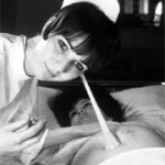
This was not a quick birth. The concept began back in 1963. After finishing each day's work at the Women's Hospital, Dusseldorf, obstetrician Konrad Hammacher spent his nights at the Medical Academy, toiling over an idea.
Arne Larsson was only 42 years old when his heart began to falter. In the late '50s it was virtually a death sentence in Sweden. But Arne saw lived to celebrate the millennium. In 1958, he had become the first patient to receive an artificial pacemaker. He had received 26 of these devices before his death in 2001, aged 86.

Bis zu 92 Prozent der so Behandelten erleben eine deutliche Beschwerden-Linderung durch die rückenmarksnahe Applizierung von Baclofen mittels einer implantierten Pumpe von Medtronic.
New research claims that nearly four million people in the UK may be unaware they are at high risk of heart disease. An University of Oxford team screened more than 71,000 people aged over 18 across England, Wales and Scotland.