Tissue Doppler Imaging

Tissue Doppler Imaging (TDI) is an emergingnon-invasive ultrasound technique that makes it possible to measure velocities at any point of the ventricular wall during the cardiac cycle. Over the last ten years, TDI has been proposed as a feasible and useful imaging tool that provides information on regional wall motion dynamics.

Fig. 2: Two-dimensional colour coded velocities of left atrial appendage walls....
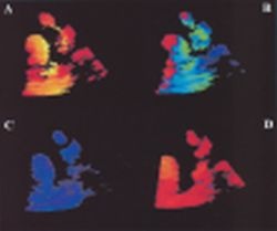
Fig. 2: Two-dimensional colour coded velocities of left atrial appendage walls. Left atrial appendage walls are depicted in lighter red (A) and blue colour (B) because of the motion of the appendage to and from the probe (contraction and relaxation). Left atrial appendage has also passive emptying and filling and the relative wall movements are coded in blue (C) and red colour (D), respectively.

This article was first published in the VISIONS, issue 1/2001, a publication of Toshiba Medical Systems

28.08.2007

Read all latest stories

Related articles

Photo

Sponsored • Computed Tomography

Mobile Imaging Solutions: Clinical Confidence Anywhere

To bridge new equipment installations in radiology, temporary high workloads, or long-term additional capacity, Canon Medical provides mobile solutions according to customers’ needs and wishes.

Photo

Video • Redesign & refresh

Canon Medical updates Aplio ultrasound systems

Canon Medical announced the commercial launch of the Aplio i-series / Prism Edition, a complete redesign of its premium ultrasound series. In addition, Aplio a-series, Canon Medical’s…

Photo

Sponsored • All-in-one imaging trailer

Mobile solutions for the COVID-19 frontline

With public health issues continuing to make daily mainstream news headlines across the world, it is clear how much change the healthcare environment is going through. Not only are there existing…

Related products

Subscribe to Newsletter