Automatic Quantification of Left-ventricular Function

The evaluation of the size and function of the left ventricle in patients with suspected heart disease is a central diagnostic problem. In contrast to other methods of evaluating left-ventricular function (e.g., angiocardiography, right-sided heart catheterization, radionuclide ventriculography or MRI), transthoracic echocardiography is a widespread and readily available procedure that is not stressful for the patient and does involve exposure to a contrast medium or to radiation.

Fig. 2: The ventricular contour is fit between the marked reference points...
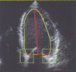
Fig. 2: The ventricular contour is fit between the marked reference points (annulus of the mitral valve and LV apex) by A-ACT (partial shape constraint contour model)

This article was first published in the VISIONS, issue 3/2002, a publication of Toshiba Medical Systems

27.08.2007

Read all latest stories

Related articles

Photo

Sponsored • Computed Tomography

Mobile Imaging Solutions: Clinical Confidence Anywhere

To bridge new equipment installations in radiology, temporary high workloads, or long-term additional capacity, Canon Medical provides mobile solutions according to customers’ needs and wishes.

Photo

Video • Redesign & refresh

Canon Medical updates Aplio ultrasound systems

Canon Medical announced the commercial launch of the Aplio i-series / Prism Edition, a complete redesign of its premium ultrasound series. In addition, Aplio a-series, Canon Medical’s…

Photo

Sponsored • All-in-one imaging trailer

Mobile solutions for the COVID-19 frontline

With public health issues continuing to make daily mainstream news headlines across the world, it is clear how much change the healthcare environment is going through. Not only are there existing…

Related products

Subscribe to Newsletter