Healthcare economics
Research using an analytical health economics model has suggested the current system of screening within the UK’s National Health Service (NHS) is only moderately likely to be cost effective.


Research using an analytical health economics model has suggested the current system of screening within the UK’s National Health Service (NHS) is only moderately likely to be cost effective.

With the help of a commercially available CAD (computer-assisted diagnosis) programme, MRI can provide prognostic data on the development of distant metastases in the further course of breast cancer.
Ultrasound may be used during breast conservation surgery, to locate tumour lesions or to place localising wires; it can also guide a lumpectomy and perform a specimen exam to ensure a lesion has been excised and to evaluate surgical margins

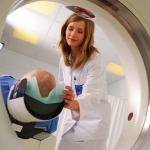
Researchers in Germany have suggested that, for certain patients, newly developed coronary CT angiography techniques can provide good quality images with very low dose radiation.
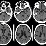
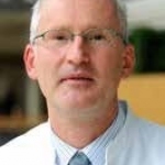
‘We finally have tools to non-invasively study the human brain in normal subjects and diseased patients,’ says Professor Stefan Sunaert, Head of Translational MRI at the Department of Imaging & Pathology, Leuven University Hospital (Belgium)
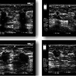
Over the decades of breast imaging numerous studies have shown that radiation free and inexpensive ultrasound can detect some subtle cancers not visible on a mammography exam.

Remote US examinations is not science-fiction; they are now available for real-time diagnostics.

The charity Cancer Research UK reports that the number of breast cancer diagnoses in under 50-year-old women each year in the UK has exceeded 10,000 for the first time.

Self-guided study of more than 6,000 ultrasound exams are at your fingertips thanks to an eight-year voluntary effort by a Dutch radiologist and technical support provided by Hitachi-Aloka.
COCIR, the European trade association covering medical diagnostics, welcomes the adoption yesterday by European Council of the revised Directive on Protecting Workers from Exposure to Electromagnetic Fields, also known as the EMF Directive.
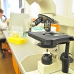
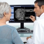
Evolving technologies and market forces reveal that digital pathology is poised to radically affect the daily workflow and activities of pathologists and diagnostic laboratories
Medical Equipment Solutions and Applications (MESA) and Euromedic International have agreed to extend their current diagnostic imaging service and maintenance partnership covering Euromedic’s Tier 1 (MRI, CT, PET-CT, Gamma Camera and Angio) and Tier 2 (mammography, ultrasound and other general X-ray) systems.
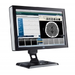
Healthcare imaging expert Barco has signed a worldwide agreement with Ventana Medical Systems, Inc., a member of the Roche Group, to provide its leading diagnostic and clinical review display systems for use with the Ventana Virtuoso image and workflow management system, offering a best-in-class, turnkey image viewing solution.
At this year’s ECR the first of two Management in Radiology (MIR) sessions addressed the issue of innovation management and future challenges.
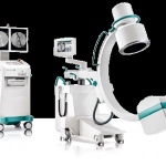
Taking a comprehensive approach to reducing dose, each link in the imaging chain was reengineered to deliver a superior clinical image with Ziehm Vision mobile C-arms. The importance of imaging for interventional radiologists is clear the moment you step into the operating theatre.
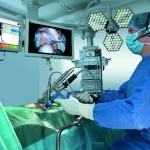
TRUMPF’s youngest child exceeds all expectations. Since November 2012, the active assistance system ViKY has been a new member in the TRUMPF product portfolio. As many as 36 orders from Europe and the Middle East has the medical technology company received so far - five of them from Turkish hospitals solely.
The future will be aesthetic or, put another way, Art meets Science. With this motto, the 43rd Congress of the German Society for Endoscopy and Imaging Procedures e.V., jointly held in Munich with six other specialist associations, demonstrated that aesthetic means the brilliance of images generated by the latest generation of X-ray, CT, MRI and ultrasound equipment.

“We finally have tools to non-invasively study the human brain in normal subjects and diseased patients,” says Professor Stefan Sunaert, Head of Translational MRI at the Department of Imaging & Pathology at Leuven University Hospital (Belgium).

ESR President Gabriel Krestin introduced a major theme for the society's congress with personalized radiology. "There is nothing more personal in healthcare than a medical image," he told fellow radiologists.
It is an every-day occurrence in any emergency department: patients presenting with severe flank pain. In roughly 50 percent of these cases, the pain is caused by a stone. 15 percent of all men and six percent of all women suffer from stones in kidney, ureter, bladder or urethra at least once in their lifetime
Management in Radiology (MIR): this ESR subcommittee is dedicated to management topics, developments in eHealth, and major trends in the discipline.
A UK hospital is assessing trauma patients by taking them directly for CT scans rather than to the A&E department. Piloted at King’s College Hospital, this new approach to assessing patients with life-threatening injuries aims to speed up diagnosis by conducting CT simultaneously with patient resuscitation and stabilisation.
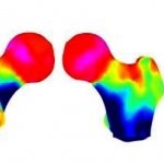
DMS (Diagnostic Medical Systems) is thrilled to unveil, for the occasion of the ECR 2013, the newest breakthrough in bone health management: 3D-DXA.
It is said a picture is worth 1,000 words. Advanced medical imaging, such as 3D views of the heart or brain have certainly proven the value of this statement by advancing our understanding of the complex biological structures and processes of disease.
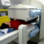
MRI has become the gold standard for many indications in cardiac imaging, apart from imaging the coronary arteries. For function and morphology assessment, MRI is the leading technology. A further advance into as yet unknown territory is myocardial imaging aided by one of the first integrated 3-Tesla PET/MR systems currently used at the Institute of Radiology, Essen University Hospital,…