Modern transducers enhance ductal echography
Too old for technological innovation? Never!
Ductal echography, meaning imaging the ducts of the female breast, is an established ultrasound procedure that may offer unexpected new insights due to technological innovations.

So explains Deputy Senior Consultant Dr Zanetti-Dällenbach, who is convinced that radial ductal echography – or radial ultrasound – can provide relevant information in breast assessment.
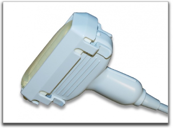
‘During a conventional ultrasound scan the investigator examines the breast in a meandering movement, which means the section is coincidental rather than anatomy specific. The new and large transducers Hitachi developed for Real-time Tissue Elastography, HI-RTE for short, in radial ultrasound, allow the examination of the breast with a circular movement. ‘In a small breast, one circle is sufficient, larger breasts require two circles.’ The major advantage of radial ultrasound is the fact that not only breast tissue but also breast anatomy is well visualised. The duct and the lobules can be assessed in longitudinal sections, which offers additional diagnostic information. Dr Zanetti- Dällenbach: ‘The visualisation of the anatomy is really superb and some experts maintain that radial ultrasound is the better method to detect small tumours. However, more importantly, it enables the investigator to evaluate better whether a detected lesion is relevant or not. Do I see a cyst protruding from the duct or is it rather a thickened cyst that is not yet anatomically suspicious? This is the type of question radial ultrasound can answer with the help of the large transducers.’
Thus the newly developed large transducers cast an entirely new light on the tried and true method of ductal echography. This may well lead to a reassessment of the method’s usability and value. It remains to be seen whether and what advantages the new approach offers because further research is needed.
However, she is excited about the new possibilities this procedure opens up and uses it regularly as a supplementary examination and as a comparison to conventional ultrasound. Nonetheless, she cautions: ‘Radial ultrasound does require experience. Moving the transducer smoothly around the nipple while looking at the monitor is not so easy.’ For patients, HI-RTE for ductal echography is far more comfortable, because the water bag softens the pressure applied to the breast. Despite the pouch, high quality images are acquired.
The large transducers used for ductal echography were especially developed by Hitachi for its Real-time Tissue Elastography application. *Hitachi Real-time Tissue Elastography (HI-RTE) is an innovative modality to assess tissue elasticity. The system has a complementary diagnostic role in B-mode imaging, providing further criteria for the characterisation of lesions. Second generation HI-RTE includes the Strain Ratio, a quantitative method for analysing the elastography score.
04.09.2012